
Acute Kidney Injury
Also known as: AKI, Acute renal failure
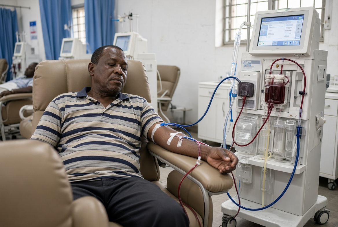
Acute kidney injury is a sudden decline in renal function, occurring over hours to days, causing accumulation of nitrogenous waste products and disturbance of fluid, electrolyte, and acid base homeostasis. It is defined and staged using the KDIGO criteria, based on the rise in serum creatinine from baseline or the fall in urine output over a defined period.
Causes are classified anatomically into three categories, and this classification drives the initial diagnostic approach.
Pre-renal injury results from reduced renal perfusion without intrinsic kidney damage, and is the most common category overall. Causes include hypovolemia (hemorrhage, dehydration, gastrointestinal losses), reduced effective circulating volume despite normal or increased total body fluid (heart failure, cirrhosis with ascites), sepsis and other causes of systemic vasodilation, and renal artery stenosis or other renal vascular compromise.
Intrinsic renal injury reflects direct damage to the renal parenchyma itself. Acute tubular necrosis, most commonly from prolonged pre-renal insult or nephrotoxin exposure, is the most common intrinsic cause. Other intrinsic causes include glomerulonephritis (see Nephritic Syndrome entry), acute interstitial nephritis (frequently drug induced, classically from NSAIDs, penicillins, or proton pump inhibitors), and vascular causes such as vasculitis or thrombotic microangiopathy.
Post-renal injury results from obstruction to urine outflow anywhere along the urinary tract, including benign prostatic hyperplasia, nephrolithiasis (particularly if bilateral or affecting a single functioning kidney), pelvic malignancy, and retroperitoneal fibrosis.
