
Haemochromatosis
Also known as: Hemochromatosis, Iron overload
Disorder of iron overload, most commonly hereditary (autosomal recessive, predominantly caused by HFE gene mutations, particularly C282Y homozygosity, most prevalent in populations of Northern European descent), causing inappropriately increased intestinal iron absorption and progressive deposition in the liver, heart, pancreas, joints, skin, and pituitary gland.
Secondary iron overload occurs from repeated blood transfusion (see Sickle Cell Disease and Leukemia entries) or, less commonly, from excessive iron intake or ineffective erythropoiesis in conditions such as thalassemia.
Early disease is often asymptomatic or presents with nonspecific fatigue and arthralgia (classically affecting the second and third metacarpophalangeal joints, sometimes termed the iron fist sign).
As iron accumulation progresses, organ specific features emerge: hepatomegaly progressing to cirrhosis and a markedly increased risk of hepatocellular carcinoma even in the absence of established cirrhosis, distinguishing this cause from most other cirrhotic etiologies; bronze or slate grey skin pigmentation (from both iron deposition and increased melanin); diabetes mellitus (from pancreatic iron deposition, historically termed bronze diabetes when combined with the skin pigmentation and cirrhosis); cardiomyopathy (restrictive or dilated, with associated arrhythmia) and heart failure; hypogonadism from pituitary iron deposition (reduced libido, erectile dysfunction, amenorrhea); and arthropathy as above.
Transferrin saturation is the most sensitive initial screening test, elevated (generally above 45%) even before ferritin rises significantly, and is the preferred first test in suspected cases or family screening.
Serum ferritin is elevated but, as an acute phase reactant, can be raised by inflammation, infection, alcohol use, or metabolic syndrome independent of true iron overload, so it should always be interpreted alongside transferrin saturation and clinical context rather than in isolation.
Genetic testing for HFE mutations (C282Y and H63D) confirms hereditary hemochromatosis once biochemical iron overload is identified.
Liver biopsy with hepatic iron index, once the primary confirmatory test, is now reserved for cases where genetic testing is negative or equivocal despite biochemical evidence of overload, or to assess the degree of fibrosis in patients with very high ferritin (generally above 1000 micrograms per liter) who are at higher risk of established cirrhosis.
MRI can quantify hepatic and cardiac iron content non-invasively and is increasingly used both for diagnosis and for monitoring treatment response, particularly in transfusion related secondary overload.
Differentials include other causes of chronic liver disease with elevated ferritin (alcohol related liver disease, MASLD, viral hepatitis, all of which can cause a reactive ferritin elevation without true iron overload), and secondary iron overload from transfusion or ineffective erythropoiesis.
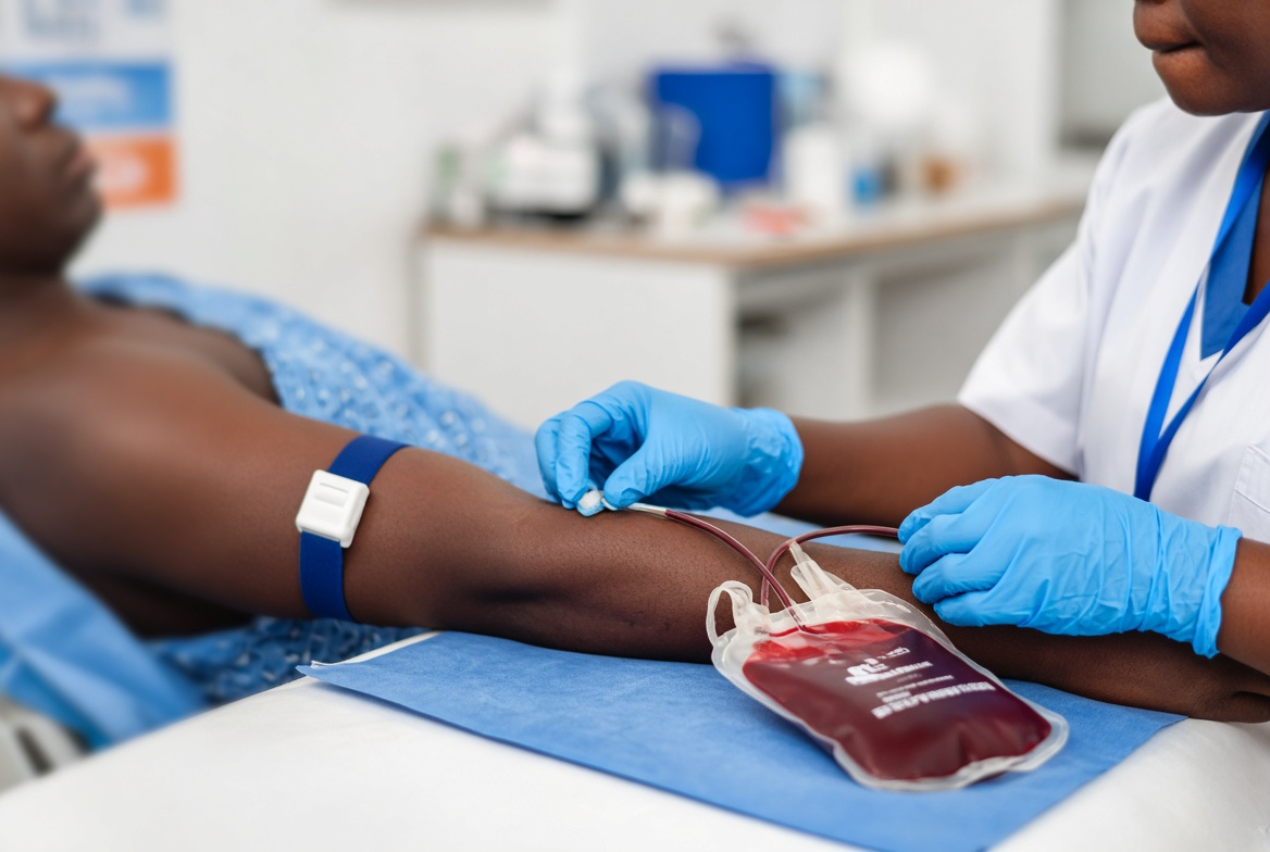
Therapeutic phlebotomy is the mainstay of treatment for hereditary hemochromatosis: weekly or twice weekly venesection of approximately 450 to 500 mL initially, continued until ferritin falls to the target range (generally 50 to 100 micrograms per liter), followed by maintenance phlebotomy every few months thereafter to keep iron stores within target range lifelong.
Treatment initiated before the onset of cirrhosis or diabetes can prevent these complications entirely and restores normal life expectancy, which is why early identification through family screening of first degree relatives once an index case is diagnosed is a central part of management.
Dietary advice includes avoiding iron supplements and excessive vitamin C intake (which enhances iron absorption), and moderating alcohol intake given the compounding effect on liver injury, though strict dietary iron restriction is not generally necessary once phlebotomy is established.
Iron chelation therapy (deferasirox, deferoxamine, or deferiprone) is reserved for secondary iron overload where phlebotomy is not feasible due to underlying anemia, such as in transfusion dependent conditions.
Once cirrhosis has developed, hepatocellular carcinoma surveillance (six monthly ultrasound with or without AFP) continues indefinitely even after iron stores are normalized by treatment, since established cirrhosis carries ongoing cancer risk regardless of subsequent iron status.
Referral: hepatology or gastroenterology for diagnosis confirmation and management; hematology for phlebotomy program coordination; genetic counseling and screening for first degree relatives once an index case is confirmed.
That completes the GI/Hepatobiliary group (items 1 to 17). Send the word when you want the next group (Cardiology and Respiratory, items 18 to 28).

