

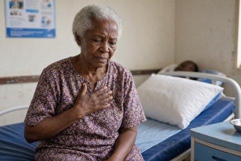

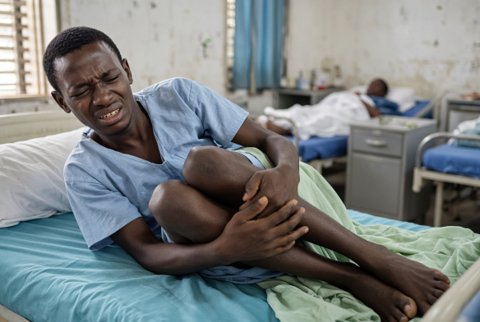








Bronchiectasis
Bronchiectasis is permanent, abnormal dilatation of the bronchi with chronic inflammation, impaired mucociliary clearance and recurrent infection. It follows a vicious cycle of infection, inflammation and airway damage.
Prevalence is about 100 to 500 per 100,000 in adults in high income regions and rising, with a female predominance. Higher rates occur in Indigenous and low income populations, often after childhood infection.
Causes: post infectious (tuberculosis, pertussis, measles, severe pneumonia), cystic fibrosis, primary ciliary dyskinesia, immunodeficiency (common variable immunodeficiency, X linked agammaglobulinaemia), allergic bronchopulmonary aspergillosis, COPD and asthma overlap, rheumatoid arthritis, inflammatory bowel disease, alpha 1 antitrypsin deficiency, aspiration, foreign body or tumour obstruction, Mounier-Kuhn syndrome, yellow nail syndrome, Young syndrome. Idiopathic in 30 to 50%.
Patterns: cylindrical, varicose, cystic. Upper lobe predominance suggests cystic fibrosis, ABPA or tuberculosis. Lower lobe suggests post infectious or aspiration. Middle lobe and lingula suggests nontuberculous mycobacteria (Lady Windermere syndrome).
Pathogens: Haemophilus influenzae, Pseudomonas aeruginosa (marker of severity), Staphylococcus aureus, Streptococcus pneumoniae, Moraxella, nontuberculous mycobacteria, Aspergillus.
Severity: Bronchiectasis Severity Index (age, BMI, FEV1, hospitalisation, exacerbations, MRC dyspnoea, Pseudomonas colonisation, other colonisation, radiological extent) and FACED score.
Complications: haemoptysis, recurrent exacerbations, respiratory failure, cor pulmonale, amyloidosis, brain abscess, mortality about 10 to 20% at 5 years.
