










DNA Viruses: Herpesviruses
Herpesviruses are large, enveloped, double-stranded DNA viruses that replicate within the host cell nucleus using a combination of host and virus-encoded enzymes. The family's defining biological property, shared by every member in this group, is the capacity to establish lifelong latency in a specific, characteristic host cell type after primary infection, with the potential to reactivate later in life, often repeatedly, whenever host immune surveillance is reduced.
Eight herpesviruses are recognised as human pathogens. Herpes simplex virus 1 and 2, varicella zoster virus, Epstein-Barr virus, cytomegalovirus, human herpesvirus 6 and 7, and human herpesvirus 8. Each establishes latency in a different cell type, and recognising that cell type is the single most useful way to predict both an individual virus's reactivation disease pattern and its diagnostic approach.
Genome. Double-stranded DNA. Envelope. Present.
Transmission. HSV-1 is transmitted predominantly by direct contact with oral secretions or lesions, classically acquired in childhood through nonsexual contact. HSV-2 is transmitted predominantly through sexual contact. This distinction is no longer absolute in practice. Oral-genital contact has made HSV-1 an increasingly common cause of genital disease, and HSV-2 can, less commonly, cause oral disease.
Latency. Both viruses establish latency within sensory neurons of dorsal root or cranial nerve ganglia. HSV-1 characteristically establishes latency in the trigeminal ganglion, reflecting its typical site of primary oral infection. HSV-2 characteristically establishes latency in the sacral dorsal root ganglia, reflecting its typical site of primary genital infection. Reactivated virus travels back down the sensory nerve axon to produce recurrent lesions at or near the original site of primary infection.
Major disease. Primary HSV-1 infection is most often asymptomatic or mild. A recognised presentation in young children is gingivostomatitis: painful vesicular and ulcerative lesions of the oral mucosa and gingiva, accompanied by fever and lymphadenopathy. Recurrent HSV-1 disease presents as herpes labialis, the common cold sore, typically preceded by a prodromal tingling or burning sensation before vesicle formation.
Primary and recurrent HSV-2 infection produces genital herpes: painful grouped vesicles on an erythematous base progressing to ulceration, with regional lymphadenopathy and, in primary infection, systemic symptoms. Recurrent episodes are generally milder and shorter than the primary episode, since partial immunity blunts subsequent reactivation severity.
Both viruses can cause disease well beyond their typical mucocutaneous presentation.
- Herpetic whitlow is HSV infection of a finger, classically an occupational exposure in healthcare workers before the routine use of gloves.
- Herpes gladiatorum is cutaneous HSV spread through skin-to-skin contact in contact sports such as wrestling.
- Ocular HSV infection (herpes keratitis) is a leading infectious cause of corneal blindness in high-income countries. It characteristically produces a dendritic corneal ulcer on fluorescein staining.
Two presentations carry particular severity.
- Herpes simplex encephalitis is most often caused by HSV-1. It characteristically localises to the temporal lobes, producing fever, altered consciousness, focal neurological signs, and seizures. It carries substantial mortality and neurological morbidity if treatment is delayed.
- Neonatal herpes results from transmission during vaginal delivery in a mother with active genital HSV shedding. The risk is highest when the mother acquires a new, primary genital infection late in pregnancy, since she has not yet developed protective antibody to pass to the neonate.
- Neonatal disease ranges from localised skin, eye, and mouth involvement to disseminated disease or encephalitis. The latter two carry high mortality and morbidity without prompt treatment.
Diagnosis. PCR of lesion swab or, for encephalitis, cerebrospinal fluid, is the diagnostic method of choice given its superior sensitivity over viral culture. The Tzanck smear shows multinucleated giant cells on a scraping from the base of a vesicle. It is a rapid but nonspecific bedside test that cannot distinguish HSV from VZV. Serology has limited value in acute diagnosis. It can establish past exposure or, in specific circumstances, help assess neonatal transmission risk based on maternal antibody status.
Genome. Double-stranded DNA. Envelope. Present.
Transmission. Respiratory droplets and direct contact with vesicle fluid. VZV is highly contagious, and airborne spread contributes meaningfully to transmission, particularly in disseminated zoster.
Latency. VZV establishes latency within dorsal root and cranial nerve sensory ganglia following primary infection, in the same general neuronal compartment as HSV. It is distributed across many ganglia rather than concentrated at one typical site, since primary varicella produces widespread viraemic seeding of sensory ganglia throughout the body.
Major disease. Primary infection causes varicella, chickenpox: a generalised vesicular rash appearing in successive crops over several days. Lesions at different stages (macule, papule, vesicle, crust) are visible simultaneously on the same patient. This distinguishes it from the more uniform, single-crop rash of smallpox historically and from the cephalocaudal, single-wave spread of measles. Fever and malaise accompany the rash.
Disease is generally mild in healthy children but tends to be more severe in adults, in pregnancy, and in immunocompromised hosts. There is a recognised risk of primary varicella pneumonia, particularly in adult and pregnant patients.
Reactivation produces herpes zoster, shingles: a painful, vesicular rash confined to a single dermatome, or occasionally two adjacent dermatomes. This reflects reactivation within the single sensory ganglion where latency was established at that anatomical level. Pain frequently precedes the rash by one to several days.
Complications of zoster:
- Postherpetic neuralgia is persistent pain in the affected dermatome lasting beyond rash resolution. It is the most common complication and becomes substantially more likely with increasing patient age.
- Disseminated zoster involves multiple dermatomes or presents with a widespread vesicular rash resembling primary varicella. It is a marker of significant underlying immunosuppression.
- Zoster involving the ophthalmic division of the trigeminal nerve risks corneal involvement and vision loss and is an ophthalmological emergency.
- Zoster involving the geniculate ganglion produces Ramsay Hunt syndrome: facial nerve palsy with vesicles in the ear canal and, frequently, hearing disturbance.
Congenital varicella syndrome follows maternal primary varicella infection, most significantly in the first half of pregnancy. It produces limb hypoplasia, cutaneous scarring in a dermatomal distribution, and ocular and central nervous system abnormalities. These reflect direct fetal viral damage during a period of active organogenesis and neural development.
Diagnosis. Predominantly clinical, given the highly characteristic rash of both varicella and zoster. PCR of vesicle fluid confirms the diagnosis when needed, particularly in atypical presentations or immunocompromised patients. It has replaced the nonspecific Tzanck smear as the preferred confirmatory test where available.
Genome. Double-stranded DNA. Envelope. Present.
Transmission. Saliva, classically through kissing, giving rise to the colloquial description of the resulting illness as the kissing disease. Transmission can also occur through shared drinking vessels and other saliva-contaminated contact.
Tropism and latency. EBV infects oropharyngeal epithelial cells initially, then B lymphocytes, within which it establishes lifelong latency. The virus's capacity to directly drive proliferation of infected B cells is central both to the clinical illness it produces and to its recognised oncogenic potential.
Major disease. Primary infection in young children is frequently asymptomatic or mild. Primary infection delayed until adolescence or young adulthood classically produces infectious mononucleosis: fever, severe pharyngitis often with tonsillar exudate, and generalised lymphadenopathy, frequently with prominent posterior cervical involvement. Splenomegaly occurs in a substantial proportion of patients.
Atypical lymphocytes are a characteristic peripheral blood finding. These are reactive CD8 T cells responding to the proliferating infected B cell population.
A specific and well-recognised prescribing pitfall: amoxicillin or ampicillin given to a patient with unrecognised infectious mononucleosis frequently provokes a diffuse maculopapular rash. This does not indicate true penicillin allergy but is strongly associated with this viral infection.
Splenic rupture is an uncommon but serious complication of infectious mononucleosis. The associated splenomegaly is the basis for standard advice to avoid contact sports and heavy physical exertion for several weeks during recovery.
EBV carries a well-established oncogenic association, driving proliferation of latently infected B cells through expression of specific viral latency proteins. Recognised EBV-associated malignancies:
- Endemic Burkitt lymphoma, strongly associated with the chromosomal translocation involving the MYC oncogene. It is geographically concentrated in regions of holoendemic malaria, which is believed to contribute through chronic B cell stimulation and impaired T cell surveillance.
- Hodgkin lymphoma, in a proportion of cases.
- Nasopharyngeal carcinoma, particularly prevalent in Southeast Asia and southern China.
- Post-transplant lymphoproliferative disease, arising from uncontrolled proliferation of EBV-infected B cells in the setting of the T cell immunosuppression required to prevent transplant rejection.
Diagnosis. The heterophile antibody test (the Monospot test) detects nonspecific antibodies characteristically produced during acute EBV infection. It is the standard rapid diagnostic test, though it is frequently negative in young children and may be falsely negative in the first week of illness.
EBV-specific serology, including antibodies against viral capsid antigen and EBV nuclear antigen, allows more precise staging of infection as acute, recent, or past. PCR quantification of EBV viral load is used specifically to monitor for and diagnose post-transplant lymphoproliferative disease in transplant recipients.
Genome. Double-stranded DNA. Envelope. Present.
Transmission. A wide range of body fluids, including saliva, urine, breast milk, cervical secretions, semen, and blood, reflecting broad viral shedding across multiple sites during both primary and reactivated infection. Transmission occurs through close personal contact, sexual contact, blood transfusion, organ transplantation, breastfeeding, and transplacentally.
Tropism and latency. CMV establishes latency predominantly within cells of the myeloid lineage, monocytes and their progenitors, with reactivation occurring as these cells differentiate and disseminate to multiple organs, explaining the virus's capacity to produce disease in essentially any organ system during active infection or reactivation.
Major disease. Primary infection in immunocompetent individuals is usually asymptomatic or produces a mild, self-limited mononucleosis-like illness, generally milder than classic EBV mononucleosis and typically heterophile-antibody-negative, a useful distinguishing feature when the two are clinically confused.
Congenital CMV infection is the most common congenital viral infection and the leading infectious cause of congenital sensorineural hearing loss. It can result from either primary or, less commonly, reactivated maternal infection during pregnancy, with primary maternal infection carrying substantially higher transmission and disease risk.
Affected infants may be born with growth restriction, microcephaly, hepatosplenomegaly, thrombocytopenia, chorioretinitis, and intracranial calcifications, classically periventricular in distribution, though many congenitally infected infants are asymptomatic at birth and only later develop sensorineural hearing loss.
In immunocompromised hosts, particularly solid organ and haematopoietic stem cell transplant recipients and patients with advanced HIV infection, CMV reactivation causes serious end-organ disease. CMV retinitis, a leading cause of vision loss in advanced, untreated HIV infection, produces a characteristic fundoscopic appearance of retinal haemorrhage and necrosis.
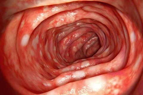
CMV colitis, oesophagitis, pneumonitis, and hepatitis are further recognised patterns of reactivation disease in this population, and CMV disease itself contributes to a broader, generalised immunosuppressive effect that increases susceptibility to other opportunistic infections in transplant recipients.
Diagnosis. PCR quantification of CMV viral load in blood is the standard method for both diagnosing active disease and monitoring transplant recipients for early reactivation, allowing pre-emptive treatment before overt end-organ disease develops. Serology distinguishes primary from past infection based on IgM and IgG status and is particularly relevant in assessing maternal infection timing during pregnancy. Tissue biopsy with characteristic owl's eye intranuclear inclusion bodies confirms organ-specific invasive disease when needed.
Genome. Double-stranded DNA. Envelope. Present.
Transmission. Saliva. Primary infection occurs almost universally in early childhood, generally between six months and two years of age, as maternal antibody wanes.
Latency. Both viruses establish latency predominantly within lymphocytes and monocytes. HHV-6 is additionally capable of chromosomal integration in a small proportion of infected individuals. This is a distinctive persistence mechanism not shared by other herpesviruses in this group.
Major disease. HHV-6, and less commonly HHV-7, causes roseola infantum (exanthem subitum). This is a febrile illness in infants and young children. Several days of high fever are followed by defervescence and the appearance of a diffuse maculopapular rash.
The rash characteristically appears only as the fever resolves. This sequence distinguishes it from most other childhood exanthems, where fever and rash occur together.
Febrile seizures are a recognised complication during the febrile phase. In most cases they reflect the high fever itself rather than direct CNS viral invasion.
In immunocompromised hosts, particularly haematopoietic stem cell transplant recipients, HHV-6 reactivation is associated with encephalitis and bone marrow suppression. This is a clinically significant complication, distinct from the mild, self-limited disease typical of primary childhood infection.
Diagnosis. Predominantly clinical for typical roseola. PCR is used to confirm reactivation disease in immunocompromised hosts, where the diagnosis is less clinically obvious than in the classic paediatric presentation.
Genome. Double-stranded DNA. Envelope. Present.
Transmission. Saliva is the principal route in regions of high endemicity. Sexual transmission, particularly among men who have sex with men, is more significant in low-prevalence populations. Transmission can also occur via organ transplantation and, less commonly, blood transfusion.
Tropism and oncogenic association. HHV-8 establishes latency within endothelial cells and B lymphocytes. It is the causative agent of Kaposi sarcoma, a vascular tumour arising from infected endothelial cells. Virus-encoded proteins promote cell proliferation and angiogenesis.
Kaposi sarcoma occurs in three settings:
- Significant immunosuppression, most notably advanced, untreated HIV infection, where it is an AIDS-defining illness.
- A classic, indolent form in elderly men of Mediterranean and Eastern European descent, independent of HIV status.
- An iatrogenic form in solid organ transplant recipients on immunosuppressive therapy.
Lesions present as violaceous cutaneous or mucosal plaques and nodules. Visceral involvement, particularly gastrointestinal and pulmonary, occurs in more advanced or aggressive disease.
HHV-8 is also causally associated with two further, less common conditions. Both occur predominantly, though not exclusively, with HIV co-infection.
- Primary effusion lymphoma: a B cell lymphoma presenting as a malignant effusion without a solid tumour mass.
- Multicentric Castleman disease: a lymphoproliferative disorder producing generalised lymphadenopathy and systemic inflammatory symptoms.
Diagnosis. Kaposi sarcoma is typically diagnosed on clinical appearance combined with biopsy, which shows the characteristic spindle cell proliferation and vascular slits of the tumour. PCR and serology can confirm HHV-8 infection status but are not required for diagnosis of typical cutaneous Kaposi sarcoma.
