








Laryngeal Cancer
Malignancy of the larynx, of which over 95 percent is squamous cell carcinoma. It accounts for around a third of all head and neck cancers and is one of the few malignancies where an early symptom, hoarseness, appears while the tumour is small and highly curable.
Anatomical subsites and why they behave differently
The behaviour, presentation, spread and prognosis of laryngeal cancer are determined almost entirely by embryological anatomy.
- Supraglottis, from the tip of the epiglottis to the laryngeal ventricle, including the epiglottis, aryepiglottic folds, arytenoids and false cords. It derives from the buccopharyngeal anlage and has a rich bilateral lymphatic network draining to levels II and III bilaterally. Consequently, supraglottic tumours present late with vague symptoms and have a 30 to 50 percent rate of occult nodal metastasis at presentation, frequently bilateral. Around 30 to 35 percent of laryngeal cancers.
- Glottis, comprising the true vocal folds and the anterior and posterior commissures, extending 1 cm below the free edge. It derives from the tracheobronchial anlage and has almost no lymphatic drainage, which is the single most important anatomical fact in this disease. Glottic tumours therefore present early with hoarseness, metastasise late, and have occult nodal disease in under 5 percent for T1 lesions. Around 60 to 65 percent of laryngeal cancers, and the group with the best prognosis, with 5 year survival above 90 percent for T1.
- Subglottis, from 1 cm below the vocal folds to the inferior border of the cricoid. Lymphatic drainage is to the paratracheal and level VI nodes and to the mediastinum. Tumours present late with airway obstruction and have a poor prognosis. Under 5 percent of laryngeal cancers.
Barriers to spread
The conus elasticus and the quadrangular membrane, the thyroid and cricoid cartilages, and the anterior commissure tendon of Broyles all resist tumour spread. Broyles tendon is a weak point where the perichondrium is absent, permitting anterior spread through the thyroid cartilage, which is why anterior commissure involvement upstages the tumour and reduces the success of transoral surgery and radiotherapy.
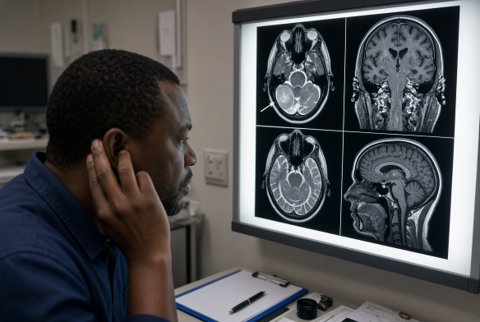
The paraglottic and pre epiglottic spaces are fat filled compartments that permit submucosal spread with a deceptively normal mucosal surface. Pre epiglottic space invasion classifies a tumour as T3 even when it appears superficially limited, and this is only detectable on imaging, which is why cross sectional imaging is required for staging even when the endoscopic appearance seems modest.
Risk factors
Tobacco and alcohol, acting synergistically, account for the great majority. Others include occupational exposure to asbestos, wood dust, nickel, sulphuric acid mist and polycyclic aromatic hydrocarbons, gastro oesophageal reflux, previous radiotherapy, human papillomavirus in a small proportion, and laryngeal papillomatosis with malignant transformation.
