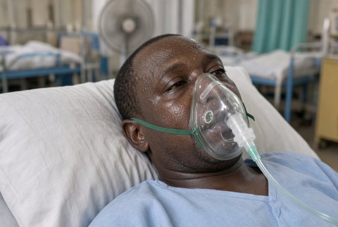
Management is predominantly supportive, following standard heart failure and arrhythmia management principles according to the severity of presentation, since no specific antiviral or targeted treatment has demonstrated consistent evidence of benefit for the common viral aetiologies.
Standard heart failure therapy is initiated according to standard heart failure guidelines once the patient is stable enough to tolerate these agents:
- an angiotensin converting enzyme inhibitor (or angiotensin receptor blocker where not tolerated)
- a beta blocker, introduced cautiously once haemodynamically stable
- a mineralocorticoid receptor antagonist
Diuretics are used for symptomatic fluid overload.
Fulminant myocarditis with cardiogenic shock requires management in an intensive or cardiac care unit. Give inotropic support (dobutamine, typically starting at 2.5 micrograms per kg per minute and titrated to haemodynamic response).
For the most severe presentations not responding adequately to pharmacological support, use mechanical circulatory support (intra-aortic balloon pump, or veno-arterial extracorporeal membrane oxygenation) as a bridge to recovery. In this specific fulminant presentation, the underlying inflammatory process, while acutely severe, frequently resolves substantially with adequate haemodynamic support.
Arrhythmia is managed according to standard protocols. Use temporary pacing for significant bradyarrhythmia or high grade atrioventricular block. Use standard antiarrhythmic therapy or, where indicated, consider an implantable cardioverter defibrillator for significant ventricular arrhythmia.
Device implantation decisions are generally deferred until the acute inflammatory phase has settled, given the potential for arrhythmia to resolve as myocarditis resolves.
Immunosuppression (corticosteroids, with additional agents such as azathioprine or ciclosporin in some protocols) is reserved for specific biopsy confirmed diagnoses where an immune mediated rather than viral mechanism is established. These are particularly giant cell myocarditis and cardiac sarcoidosis.
In giant cell myocarditis, aggressive combination immunosuppression is essential, given the otherwise poor prognosis of this specific entity.
Immunosuppression is not routinely used for presumed viral myocarditis. Evidence of benefit is inconsistent, and there is theoretical concern regarding impaired viral clearance in this broader population.
Immune checkpoint inhibitor associated myocarditis is an important and potentially fulminant cause requiring urgent recognition, given increasing use of these cancer therapies. Treat with immediate cessation of the causative agent together with high dose corticosteroids (methylprednisolone 1 g intravenously once daily for three days, followed by a prolonged oral taper).
Consider additional immunosuppression (mycophenolate, infliximab, or antithymocyte globulin in refractory cases) under specialist cardio-oncology guidance. Associated mortality is significant if not promptly and aggressively treated.
Restrict competitive sport and strenuous physical activity for a minimum of three to six months following diagnosis. This is standard advice, given evidence of increased arrhythmic risk during the active inflammatory phase. It is particularly relevant to exercise related sudden cardiac death in young athletes.
Return to activity is guided by resolution of symptoms, normalisation of cardiac biomarkers and ventricular function, and, where performed, resolution of inflammatory changes on follow up cardiac magnetic resonance imaging.
Long term follow up with serial echocardiography is important, given the risk of progression to chronic dilated cardiomyopathy in a proportion of patients. The risk is highest in those with persistent left ventricular dysfunction at the time of initial diagnosis.
Continue and titrate heart failure therapy according to standard chronic heart failure management principles in this group.