








Osteoporotic Vertebral Compression Fracture
An osteoporotic vertebral compression fracture is a collapse of a vertebral body, most often at the thoracolumbar junction. It follows low energy loading (a fall from standing height, lifting, a cough, or no recalled injury) in a patient with low bone strength.
It is the commonest fragility fracture, and a marker of high risk of further fractures.
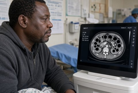
About 1.4 million occur worldwide each year; about 25% of women over 70 years and 40% of those over 80 years have at least one. Only about a third are clinically recognised, and the rest are found on imaging done for another reason.
Risk factors are age, female sex, previous fracture, low bone density, smoking, alcohol excess, low body weight and falls.
Others are glucocorticoid therapy (5 mg prednisolone daily or equivalent for over 3 months), hypogonadism, hyperparathyroidism, malabsorption and rheumatoid arthritis. Some drugs add risk (aromatase inhibitors, androgen deprivation, anticonvulsants, proton pump inhibitors long term).
After one vertebral fracture, the risk of another within the next year is about 20%. Complications are acute and chronic pain, progressive kyphosis, height loss, reduced lung function, abdominal pressure symptoms, loss of function, increased mortality (up to 20% higher than controls), nonunion with intravertebral vacuum cleft, and, rarely, neurological deficit from retropulsion.
Pathophysiology.
Loss of trabecular bone and microarchitecture reduces the strength of the vertebral body so that normal loads cause failure of the anterior column (wedge, biconcave or crush shapes). Anterior wedging increases thoracic kyphosis and moves the centre of gravity forward, which raises loads on adjacent vertebrae and causes a cascade of fractures. Pain arises from microfracture, periosteal stretch and muscle spasm.
