

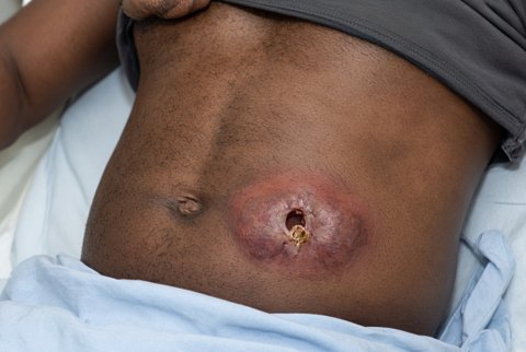










Intestinal Amoebae
Entamoeba histolytica is the only intestinal amoeba with established pathogenic potential in humans, responsible for amoebiasis, a disease ranging from asymptomatic luminal colonisation to fulminant dysentery and life-threatening extraintestinal abscess. It is transmitted by the faecal-oral route and remains a major cause of morbidity and mortality in regions with inadequate sanitation.
The trophozoite is the motile, actively feeding, invasive form. It measures roughly 10 to 60 micrometres.
It has a single nucleus with a small, centrally placed karyosome and finely beaded, evenly distributed peripheral chromatin. This nuclear pattern distinguishes it microscopically from nonpathogenic Entamoeba species and from Entamoeba dispar.
Trophozoites move by extending directional pseudopodia. They can be seen ingesting red blood cells (erythrophagocytosis). In a fresh stool specimen, this is considered strong morphological evidence of pathogenic invasive infection.
The trophozoite is fragile. It is destroyed by gastric acid and cannot survive outside the host for more than a short period. It is therefore not the transmissible stage.
The cyst is the resistant, non-motile, infective stage responsible for transmission.
Mature cysts are spherical, typically 10 to 20 micrometres in diameter. They contain up to four nuclei (a mature quadrinucleate cyst). Each nucleus has the same small central karyosome and fine peripheral chromatin pattern as the trophozoite nucleus.
Immature cysts may contain one or two nuclei and characteristic chromatoid bodies. These are rod-shaped structures with rounded, smooth ends, composed of ribonucleoprotein. They are a useful diagnostic feature on stool microscopy, particularly in early-stage cysts, where they are more prominent.
The cyst wall resists gastric acid, moderate chlorination, and environmental desiccation. Cysts survive in contaminated water and food for extended periods.
Infection follows ingestion of mature quadrinucleate cysts. The usual vehicles are faecally contaminated water, food (especially uncooked vegetables and fruit washed in contaminated water), or hands.
Excystation occurs in the small intestine, triggered by the alkaline pH and digestive enzymes. It releases a single amoeba, which undergoes nuclear division to produce eight trophozoites. These then migrate to the colon.
Mechanical vectors contribute to transmission in areas of poor sanitation. Houseflies and cockroaches carry cysts on their body surface from faeces to food.
Person-to-person transmission also occurs via the faecal-oral route in settings of close contact and poor hygiene, and via anal-oral sexual contact.
Trophozoites colonise the large intestine, principally the caecum, ascending colon, and rectosigmoid region.
In most infected individuals the organism remains a luminal commensal. It feeds on bacteria and intestinal debris without invading the mucosa, producing asymptomatic cyst passage.
In a subset of infections, trophozoites adhere to the colonic epithelium via a galactose/N-acetylgalactosamine-specific lectin. They disrupt the mucous layer and invade the mucosa, triggering the tissue-destructive phase of disease.
Invasion is mediated by amoebic proteins:
- the Gal/GalNAc lectin (adherence)
- amoebapores, pore-forming proteins that lyse host cells
- cysteine proteinases, which degrade extracellular matrix and mucins
Together they produce contact-dependent cytolysis of host epithelial and inflammatory cells.
The characteristic histopathological lesion of invasive intestinal amoebiasis is the flask-shaped ulcer. It is a narrow mucosal breach that widens as it extends through the submucosa. Trophozoites invade laterally beneath an intact or minimally disrupted surface epithelium.
This undermining pattern, narrow neck and broad base, distinguishes amoebic ulcers from the more diffuse mucosal ulceration typical of bacterial colitis.
Between ulcers, the mucosa is often relatively normal. This patchy distribution also differs from the continuous mucosal involvement seen in inflammatory bowel disease.
Extensive submucosal spread can lead to coalescence of adjacent ulcers and mucosal sloughing. In severe cases it causes full-thickness necrosis with perforation.
Symptomatic intestinal amoebiasis typically presents as amoebic colitis. Onset is gradual, over one to several weeks. This distinguishes it from the abrupt onset of bacterial dysentery.
Abdominal pain, cramping, and diarrhoea progress to dysentery: frequent, small-volume stools containing blood and mucus. Fever is little or absent in most cases. This helps distinguish it from Shigella dysentery, where fever is typically prominent. Tenesmus is common.
Severe or fulminant necrotising colitis is a recognised complication. It occurs particularly in young children, pregnant women, malnourished individuals, and those on corticosteroids. It carries high mortality and can progress to toxic megacolon and perforation.
Other recognised intestinal complications are amoeboma, a mass of granulation tissue that can mimic colonic carcinoma, and chronic non-dysenteric colitis.
The most important extraintestinal manifestation is amoebic liver abscess. Trophozoites enter mesenteric venules, travel via the portal circulation to the liver, and establish a focus of tissue necrosis, most often in the right lobe.
Liver abscess can develop without concurrent diarrhoea or a clear history of dysentery. Many patients have no active intestinal symptoms at presentation.
Classic features are right upper quadrant pain (which may radiate to the right shoulder), fever, tender hepatomegaly, and a raised right hemidiaphragm on imaging.
The abscess contains characteristic "anchovy paste" material. This is a reddish-brown, odourless fluid of necrotic liver tissue and blood. Trophozoites are typically found at the abscess wall rather than in the central necrotic material. This has practical implications for aspirate sampling if diagnostic aspiration is performed.
Complications include rupture into the pleural space or pericardium. Pericardial rupture carries particularly high mortality. Rarely, the infection disseminates to brain or other distant sites.
Stool microscopy remains a widely used initial test. On direct wet mount or after concentration, it identifies cysts (quadrinucleate, with chromatoid bodies) or trophozoites (with ingested red cells supporting a pathogenic diagnosis).
A critical limitation is that Entamoeba histolytica is morphologically identical to the nonpathogenic species Entamoeba dispar and Entamoeba moshkovskii. Microscopy alone cannot reliably distinguish pathogenic from nonpathogenic infection. This matters because Entamoeba dispar is far more prevalent worldwide and does not require treatment.
Stool antigen detection (enzyme immunoassay targeting the Gal/GalNAc lectin) provides species-specific identification of E. histolytica. It is more sensitive and specific than microscopy for distinguishing it from nonpathogenic species.
Stool PCR offers similarly high specificity and sensitivity and is increasingly used where available.
Serology (detecting antibody against E. histolytica) is particularly useful in suspected extraintestinal disease (liver abscess). Stool examination is frequently negative there, since the organism has left the intestinal lumen. Serology is positive in the large majority of true invasive amoebiasis cases. In endemic populations, a positive result cannot always distinguish current from past infection.
Imaging (ultrasound or CT) demonstrates the hepatic abscess and guides the differential from pyogenic liver abscess.
Aspiration is generally reserved for diagnostic uncertainty, large abscesses at risk of rupture, or failure to respond to medical therapy. It is not performed routinely, given the risk of the procedure and the accuracy of combined serology and imaging in typical presentations.
Several other Entamoeba species and related genera colonise the human intestine without established pathogenic potential, and correct identification prevents unnecessary treatment.
Entamoeba dispar is morphologically identical to E. histolytica but genetically and biochemically distinct, does not invade tissue, and does not cause disease; it is substantially more common globally than true E. histolytica infection, meaning that microscopy-based diagnosis without species-specific confirmation considerably overestimates pathogenic infection.
Entamoeba coli is a common nonpathogenic commensal distinguished microscopically by a larger cyst (up to 8 nuclei when mature) and a coarser, more irregularly arranged peripheral nuclear chromatin with an eccentrically placed karyosome, contrasting with the fine, even chromatin and central karyosome of E. histolytica.
Other nonpathogenic species and organisms occasionally seen on stool microscopy include Endolimax nana and Iodamoeba butschlii, both distinguished by their nuclear morphology.
Endolimax nana has a large, irregular karyosome without peripheral chromatin. Iodamoeba butschlii has a large karyosome and a characteristic prominent glycogen vacuole in the cyst that stains densely with iodine.
Recognising these as nonpathogenic prevents inappropriate treatment, though their presence indicates faecal-oral exposure and should prompt consideration of other pathogens acquired through the same route.
