










Parasitology Revision Framework
Working through the same eleven questions in a fixed order gives any parasite a complete clinical and biological profile.
- Classification (protozoan, nematode, cestode, trematode, or ectoparasite)
- Definitive host
- Intermediate host
- Vector
- Infective stage
- Diagnostic stage
- Route of transmission
- Site in the human body
- Key life cycle step
- Major pathology
- Diagnostic method
The order reflects genuine dependency rather than arbitrary sequence. Classification narrows the likely biology before any specific detail is considered.
Host and vector identification determine which transmission route is even possible. Infective and diagnostic stage identification depend on already knowing the structure of the life cycle.
Site in the body and major pathology follow directly from where the organism resides once transmission has occurred. Diagnostic method follows from all the other answers rather than standing independently.
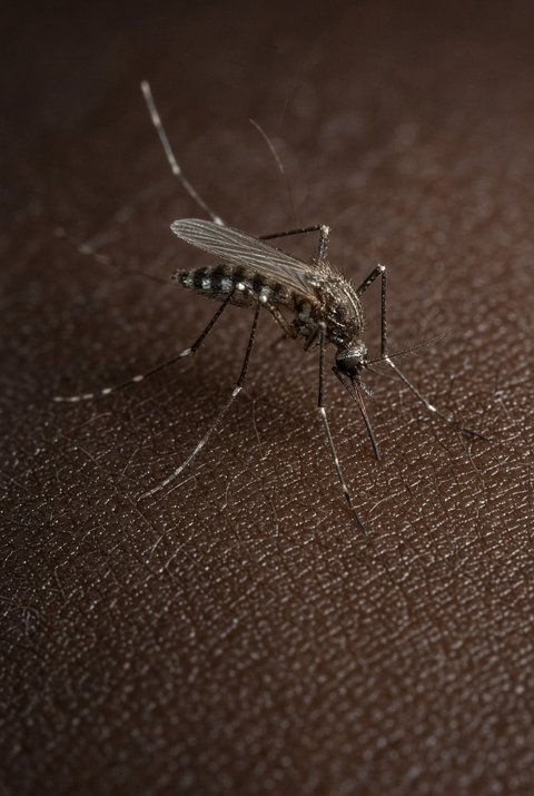
Classification: apicomplexan protozoan. Definitive host: the Anopheles mosquito, since sexual reproduction (gametogony, fertilisation, sporogony) occurs there. Intermediate host: the human, in whom only asexual replication (exo-erythrocytic and erythrocytic schizogony) occurs. Vector: female Anopheles mosquito, a true biological vector since the parasite develops within it.
Infective stage: the sporozoite, injected during the mosquito bite. Diagnostic stage: asexual erythrocytic forms, ring, trophozoite, and schizont, identified on Giemsa-stained blood film; the infective sporozoite itself is never the form sought diagnostically. Route of transmission: mosquito bite (biological vector transmission).
Site in the human body: initially the liver (clinically silent exo-erythrocytic schizogony), then red blood cells (the site of all clinical disease), with Plasmodium falciparum additionally sequestering in deep microvasculature through cytoadherence. Key life cycle step: schizont rupture, releasing merozoites and triggering both the next cycle of red cell invasion and the febrile paroxysm.
Major pathology: haemolysis, cytoadherence-driven microvascular obstruction, and, in severe disease, cerebral and placental sequestration. Diagnostic method: thick and thin blood film remains the reference standard, supported by rapid antigen testing and, where needed, PCR.
Classification: nematode. Definitive host: the human is the only host in which the parasite reaches sexual maturity; there is no separate definitive host of a different species. Intermediate host: none in the standard sense, though a free-living external generation can occur in the environment. Vector: none; transmission is by direct skin penetration, not by an arthropod.
Infective stage: the filariform (L3) larva, unique among the intestinal nematodes for being a larva rather than an egg. Diagnostic stage: rhabditiform larvae in stool, since eggs hatch within the intestinal mucosa before passage and are therefore never the form identified on routine stool examination. Route of transmission: skin penetration from contaminated soil, or, distinctively, autoinfection entirely within the same host without any new external exposure.
Site in the human body: duodenal and jejunal mucosa for the parthenogenetic adult female, with a migratory hepato-pulmonary phase preceding intestinal maturation. Key life cycle step: the capacity of rhabditiform larvae to transform into infective filariform larvae while still within the host, enabling autoinfection.
Major pathology: usually mild chronic gastrointestinal symptoms, but with potential for hyperinfection and disseminated disease, including secondary Gram-negative bacteraemia from larvae breaching the gut wall, in immunosuppressed hosts. Diagnostic method: serial stool examination with concentration techniques, serology, and, in hyperinfection, examination of sputum or other body fluids for larvae.
Classification: cestode. Definitive host: the human, harbouring the adult intestinal tapeworm. Intermediate host: the pig under normal transmission, though a human can also serve as an accidental intermediate host, giving rise to cysticercosis rather than taeniasis. Vector: none; transmission does not involve an arthropod.
Infective stage: for taeniasis, the cysticercus in undercooked pork; for cysticercosis, the egg, ingested via faecal-oral contamination or autoinfection from one's own intestinal worm. Diagnostic stage: proglottids or eggs in stool for taeniasis; imaging findings (calcified or active cysts) and serology for cysticercosis, since the larval cyst itself is not accessible to stool examination. Route of transmission: ingestion of undercooked pork (taeniasis) or ingestion of eggs (cysticercosis).
Site in the human body: small intestinal lumen for the adult worm (taeniasis); skeletal muscle, subcutaneous tissue, eye, and, most importantly, the central nervous system for the larval cysticercus (cysticercosis). Key life cycle step: the biological divergence depending on which infective stage, cysticercus versus egg, initiates infection, which determines whether the human occupies the definitive-host role or the intermediate-host role.
Major pathology: taeniasis is usually mild; cysticercosis, particularly neurocysticercosis, is the major clinical concern, with seizures typically triggered by the inflammatory response to a degenerating rather than a quiescent cyst. Diagnostic method: stool microscopy for taeniasis; neuroimaging (CT or MRI) combined with serology for neurocysticercosis.
Classification: trematode. Definitive host: the human, harbouring paired adult worms within the mesenteric venules. Intermediate host: a specific freshwater snail (Biomphalaria species), within which extensive asexual multiplication of larval stages occurs. Vector: none in the conventional sense; the snail is an intermediate host rather than a biological vector, and there is no arthropod bite involved.
Infective stage: the cercaria, the free-swimming larval form released from the infected snail, which penetrates human skin directly. Diagnostic stage: eggs (with a characteristic lateral spine for this species) identified in stool. Route of transmission: direct skin penetration during contact with contaminated freshwater, not ingestion.
Site in the human body: mesenteric venules for the adult worm pair, with trapped eggs additionally swept into the liver via portal blood flow, making the liver the principal site of chronic pathology. Key life cycle step: granuloma formation around eggs trapped in tissue, which, rather than the adult worms themselves, drives essentially all of the chronic disease.
Major pathology: periportal ("pipestem") hepatic fibrosis and presinusoidal portal hypertension, with relative preservation of hepatocyte function. Diagnostic method: stool microscopy (Kato-Katz for quantification), supported by antigen testing and, in travellers with limited exposure history, serology.
Classification: ectoparasite (arachnid mite). Definitive host: not applicable in the conventional parasitological sense; the human is simply the host on and within whose skin the entire mite life cycle occurs. Intermediate host: none. Vector: none; the mite is itself the disease-causing organism rather than a carrier of a separate pathogen.
Infective stage: the fertilised female mite, transmitted by direct skin-to-skin contact. Diagnostic stage: the mite, eggs, or faecal pellets identified microscopically from skin scraping of a burrow, or the burrow itself recognised clinically. Route of transmission: prolonged direct skin-to-skin contact, or, particularly in crusted scabies, contaminated bedding and clothing.
Site in the human body: the superficial epidermis (stratum corneum), where the female mite burrows and lays eggs, with classic sites including finger webs, wrists, and genital area.
Key life cycle step: the several-week interval between initial infestation and the onset of symptomatic pruritus, reflecting the time needed to develop a delayed-type hypersensitivity response to mite antigens, versus the far more rapid symptom onset on reinfestation in an already sensitised host.
Major pathology: intensely pruritic dermatitis from hypersensitivity to mite saliva, eggs, and faecal material, with a distinct hyperinfested crusted variant in immunocompromised or neurologically impaired hosts. Diagnostic method: clinical recognition of burrows and distribution pattern, confirmed where needed by microscopy of skin scrapings.
Applying the same eleven questions to any parasite, whether a protozoan, helminth, or ectoparasite, produces a complete and comparable profile in the same fixed structure. This allows direct comparison across organisms that differ enormously in biology, geography, and clinical presentation.
The framework forces an explicit answer to each question in turn. Gaps most often appear at the host and vector questions, where definitive and intermediate hosts are confused or a vector is assumed where transmission is actually direct.
Gaps also appear at the infective-versus-diagnostic-stage distinction. These two points of confusion recur most often across the whole of medical parasitology.
