


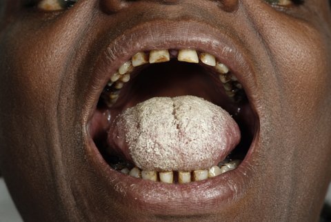









Strongyloides
Infection with Strongyloides stercoralis begins when filariform (L3) larvae, present in contaminated soil, actively penetrate intact human skin. The most common site is the soles of the feet in individuals walking barefoot on contaminated ground. This directly parallels the skin-penetration route of hookworm infection.
Larvae are motile and actively seek out and burrow through the skin. They produce a local, often transient, pruritic papular or urticarial reaction at the site of entry.
This initial cutaneous response can be mild or entirely unnoticed. This contributes to the frequent absence of a clear exposure history in patients later found to be infected, sometimes decades after their last plausible exposure.
Following skin penetration, larvae enter the venous circulation and are carried to the lungs, where they break into the alveolar spaces, ascend the bronchial tree and trachea, and are swallowed, reaching the small intestine, directly paralleling the hepato-pulmonary migration route seen in Ascaris and hookworm.
This migratory phase can produce transient respiratory symptoms (cough, wheeze, occasionally haemoptysis) and eosinophilic pulmonary infiltrates, part of the broader Loeffler-type syndrome pattern shared by migrating nematode larvae, though in many infected individuals this phase passes unnoticed or with only mild, self-limited symptoms.
Larvae that complete pulmonary migration and are swallowed mature within the mucosa of the duodenum and proximal jejunum into adult female worms.
Uniquely among the intestinal nematodes covered in this module, only parthenogenetic female worms are found within human tissue. They reproduce without the need for fertilisation by a male, a biological feature not shared by any other medically important intestinal nematode in this module.
These intestinal females produce eggs that hatch within the mucosa itself before passage, releasing rhabditiform (L1) larvae directly into the intestinal lumen. This is why rhabditiform larvae, rather than eggs, are the form identified on stool examination. It is a key distinguishing diagnostic feature from every other intestinal nematode covered in this module.
The single most clinically important and biologically distinctive feature of the Strongyloides life cycle is its capacity for autoinfection. This mechanism is absent in every other helminth of medical importance. It is central to understanding both the chronic persistence of untreated infection and the potential for life-threatening disease.
Not all rhabditiform larvae are passed out in stool to continue an external free-living or soil-based cycle. A proportion within the bowel lumen can transform directly into infective filariform (L3) larvae before leaving the host. This occurs either within the intestine or on the perianal skin (from larvae that have migrated to the perianal area in stool residue).
These filariform larvae can directly re-penetrate the colonic mucosa or perianal skin and re-enter the bloodstream. They then repeat the full migratory cycle (lungs, airway, swallowing, intestinal maturation) entirely within the same host. No external soil exposure or reinfection from the environment is needed.
This autoinfective cycle allows Strongyloides stercoralis to persist and, in effect, continually reinfect a single host indefinitely. Well-documented cases of infection persist for 30, 40, or more years after the individual last had any plausible environmental exposure (for example, decades after emigration from or military service in an endemic region).
The parasite sustains its own population within that host the entire time through repeated autoinfection cycles. These run at a low, chronic, often clinically silent level, generally held in check by an intact host immune response.
Hyperinfection syndrome is a marked acceleration of the normal autoinfective cycle. It occurs when host immune control over the rate of larval transformation and reinvasion is lost or substantially weakened.
The result is a dramatically increased larval burden confined to the organs normally involved in the migratory cycle (gastrointestinal tract, lungs, and skin), without dissemination to other organ systems.
Clinically, the usual features of each stage of the migratory cycle intensify, occurring simultaneously and severely:
- abdominal pain, nausea, vomiting, and diarrhoea (sometimes with ileus or gastrointestinal bleeding)
- respiratory symptoms including cough, dyspnoea, and wheeze, and in severe cases respiratory failure and diffuse pulmonary infiltrates or haemorrhage
- recurrence or intensification of the perianal/cutaneous larva currens rash
Larva currens is a rapidly migrating serpiginous urticarial track, distinct from cutaneous larva migrans. It can advance several centimetres per hour, reflecting the underlying rapid larval movement through the dermis.
Disseminated strongyloidiasis is a further, more severe escalation. Larvae migrate beyond the organs of the normal life cycle to invade essentially any organ system, including the central nervous system, liver, kidneys, and heart. It typically follows unrecognised or inadequately treated hyperinfection.
A highly characteristic and dangerous complication is translocation of enteric Gram-negative bacteria (and occasionally other organisms) from the gut into the bloodstream. The migrating larvae carry them directly, physically breaching the intestinal mucosal barrier during their invasive passage.
This produces Gram-negative bacteraemia and sepsis. If larvae reach the meninges, it produces polymicrobial or Gram-negative meningitis. This secondary complication can dominate the clinical picture. If the underlying Strongyloides infection is not recognised and specifically treated, it will recur and can prove fatal even if the bacterial sepsis is treated in isolation.
Mortality in disseminated strongyloidiasis and severe hyperinfection is substantial, reported in a large proportion of cases even with treatment. This reflects the severity of the underlying parasitic burden. It also reflects the frequent diagnostic delay when the parasitic infection is not considered in a patient presenting primarily with sepsis, respiratory failure, or gastrointestinal symptoms.
Loss of adequate host immune control over the autoinfective cycle precipitates hyperinfection or disseminated disease. It is most strongly and classically associated with corticosteroid therapy, even at relatively modest doses and for relatively brief courses.
This is thought to act through suppression of the Th2-type immune response (particularly interleukin-5-driven eosinophil activity) that normally restrains larval transformation and migration.
The association is sufficiently strong and well established that screening for Strongyloides infection (or empirical treatment) is specifically recommended. It applies to patients from, or with plausible exposure to, endemic areas who are about to start significant corticosteroid therapy, particularly for prolonged courses.
Other recognised risk factors for hyperinfection and dissemination:
- HTLV-1 co-infection, which appears to specifically impair the Th2 response central to Strongyloides control, more so than many other forms of immunosuppression
- HIV, a comparatively less potent risk factor for hyperinfection than might be expected
- solid organ transplantation and other immunosuppressive regimens
- haematological malignancy
- malnutrition
- any other condition producing significant impairment of cell-mediated or Th2-type immunity
This risk profile is the central reason Strongyloides receives dedicated emphasis distinct from the other intestinal nematodes. Ascaris, Trichuris, and hookworm produce disease proportional to worm burden acquired from the environment and cannot suddenly worsen without new exposure.
Strongyloides infection, however longstanding, mild, and previously asymptomatic, carries a latent potential for sudden, severe, and life-threatening escalation. A change in the host's immune status alone can trigger it, with no new external exposure required.
Strongyloides stercoralis is diagnosed by identifying rhabditiform larvae on stool microscopy, not eggs, since egg hatching occurs within the intestinal mucosa prior to passage. Filariform larvae are occasionally seen in hyperinfection, when larval burden and transit rate are greatly increased.
This has direct practical diagnostic consequences. Larval output in chronic, low-grade infection is often low and intermittent, so a single stool examination has poor sensitivity. The following improve sensitivity:
- repeated stool examinations
- concentration techniques (such as the Baermann technique or agar plate culture, both of which exploit larval motility to improve detection)
- serology, useful for chronic infection given the persistent antigenic stimulation from ongoing autoinfection, though with limited ability to distinguish active from past treated infection and with potential cross-reactivity with other helminth infections
- stool PCR
This is particularly important given the consequences of missing chronic infection in a patient who may later require immunosuppressive therapy.
Larval output in stool is frequently low and intermittent, and missed diagnosis before immunosuppression can be fatal. A diagnostic strategy for Strongyloides therefore typically combines multiple modalities rather than a single stool examination:
- serial stool examinations (ideally three or more samples on separate days) with a concentration technique
- serology in patients from endemic areas or with risk factors (accepting its limitations in distinguishing active from resolved infection)
- where hyperinfection or dissemination is suspected, examination of sputum, bronchoalveolar lavage fluid, or other body fluids for larvae
Larvae can be strikingly abundant in these severe presentations, given the massively increased larval burden and dissemination beyond the normal gastrointestinal/pulmonary migratory route.
Eosinophilia is a useful supportive clue in chronic, uncomplicated strongyloidiasis. It is frequently blunted, reduced, or entirely absent in hyperinfection and disseminated disease, since the corticosteroid or other immunosuppressive state driving the complication itself suppresses eosinophil production and function.
The absence of eosinophilia must not be used to reassuringly exclude severe strongyloidiasis in an appropriately at-risk, unwell patient. This can appear counterintuitive relative to the eosinophilia associated with most other tissue-migratory helminth infections.
