










Phaeohyphomycosis
Also known as: Dematiaceous Fungal Infection
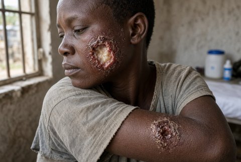
Phaeohyphomycosis is a heterogeneous group of infections caused by dematiaceous (melanised) fungi that appear in tissue as pigmented, septate hyphae, pseudohyphae or yeast like cells. The name describes the tissue form, not one organism or one syndrome. Dozens of genera and species are implicated, and the clinical range runs from a subcutaneous cyst to fatal cerebral abscess.
Phaeohyphomycosis is defined by exclusion. It is not chromoblastomycosis (muriform cells), not eumycetoma (grains), and not the superficial keratin restricted pigmented infections, tinea nigra and black piedra.
Epidemiology:
- Worldwide, with most cases from tropical and subtropical regions and from warm, humid environments.
- Subcutaneous disease follows splinter or thorn injury, usually in immunocompetent people.
- Invasive disease occurs mainly in solid organ and stem cell transplant recipients, in patients on corticosteroids, in haematological malignancy and in CARD9 deficiency. Cerebral disease also occurs in immunocompetent hosts.
- Healthcare associated clusters have followed contaminated injectable products.
All are Ascomycota. The commonest agents belong to Chaetothyriales and Pleosporales.
| Organism | Order | Typical disease | Notes |
|---|---|---|---|
| Exophiala jeanselmei complex, Exophiala spinifera | Chaetothyriales | Subcutaneous cyst, mycetoma like lesions, catheter related fungaemia | Soil, wood, water; cyst in transplant recipients |
| Exophiala dermatitidis | Chaetothyriales | Cerebral disease in young people in East Asia; airway colonisation in cystic fibrosis; fungaemia | Tolerates heat and humidity; found in steam baths, dishwashers and water systems |
| Cladophialophora bantiana | Chaetothyriales | Cerebral abscess | Neurotropic; frequently in immunocompetent patients; high mortality |
| Rhinocladiella mackenziei | Chaetothyriales | Cerebral abscess | Middle East, mainly the Arabian peninsula; very poor prognosis |
| Fonsecaea monophora | Chaetothyriales | Subcutaneous and cerebral disease | Also an agent of chromoblastomycosis |
| Verruconis gallopava (formerly Ochroconis gallopava) | Venturiales | Cerebral abscess, pulmonary nodules | Thermotolerant; poultry litter and self heating organic matter; transplant recipients |
| Curvularia species | Pleosporales | Allergic sinusitis, invasive sinusitis, keratitis, cutaneous and disseminated disease | Plant pathogen; humid regions |
| Bipolaris species | Pleosporales | Allergic fungal rhinosinusitis, invasive sinusitis, keratitis | Grasses and soil |
| Exserohilum rostratum | Pleosporales | Keratitis, sinusitis; meningitis and spinal infection in the 2012 outbreak from contaminated epidural methylprednisolone | Tropical and subtropical plant pathogen |
| Alternaria species | Pleosporales | Cutaneous and subcutaneous nodules in transplant recipients, sinusitis, keratitis, allergy | Ubiquitous airborne mould |
| Phialophora species | Chaetothyriales | Subcutaneous cysts, keratitis | Also a cause of chromoblastomycosis |
| Neoscytalidium dimidiatum | Botryosphaeriales | Nail and skin infection mimicking dermatophytosis; invasive disease rare | Common in the tropics |
| Cladosporium, Hortaea, Nigrograna and other genera | Various | Sporadic cutaneous and invasive disease | Many are environmental contaminants |
Cladophialophora bantiana has also been called Xylohypha bantiana and Cladosporium trichoides. The older name Wangiella dermatitidis is the former name of Exophiala dermatitidis.
Melanin in the cell wall is the common biological feature. It gives colonies their olive, brown or black colour and the hyphae a brown hue in tissue. It protects against oxidative killing and phagocytosis, and reduces susceptibility to amphotericin B and some other antifungals by binding drug.
Tissue forms:
- Brown, septate, branching hyphae 2 to 6 micrometres wide, often irregular, distorted, beaded or with swollen cells.
- Pseudohyphae and yeast like cells, sometimes budding, in the same lesion.
- Thick walled, rounded cells in cysts.
- Muriform cells are absent. Their presence changes the diagnosis to chromoblastomycosis.
Pigment is visible on haematoxylin and eosin when melanin is abundant. In lightly pigmented hyphae, Fontana Masson staining demonstrates melanin and separates dematiaceous hyphae from hyaline hyphae of Aspergillus, Fusarium and Scedosporium.
Entry is by implantation (splinters, thorns, trauma, contaminated injectables and surgical sites), by inhalation of conidia (sinus, lung and brain), and through intravascular devices.
- Subcutaneous disease: the fungus persists in the dermis and subcutis and produces a cystic, granulomatous and suppurative reaction around pigmented hyphae and fragments of the implanted vegetable material.
- Sinus disease: allergic forms are a Th2 and IgE mediated response to fungal antigen in the sinus. Invasive forms occur with neutropenia, diabetes and corticosteroid use.
- Cerebral disease: inhaled or haematogenous conidia reach the brain, and the fungi are neurotropic. The disease occurs in immunocompetent hosts too, with abscess formation.
- Immune control: neutrophils, macrophages and Th1 and Th17 responses through the Dectin and CARD9 pathway. CARD9 deficiency produces severe, extensive or disseminated phaeohyphomycosis, notably with Exophiala dermatitidis and Phialophora verrucosa, with cerebral, lymph node and visceral involvement and a high relapse rate.
Risk factors: solid organ transplantation, haematopoietic stem cell transplantation, corticosteroid therapy, haematological malignancy, neutropenia, diabetes mellitus, cystic fibrosis, intravascular devices, peritoneal dialysis, contaminated injectable products, trauma, and agricultural or outdoor exposure.
| Form | Features | Usual organisms |
|---|---|---|
| Subcutaneous phaeohyphomycotic cyst | Solitary, firm to fluctuant, painless nodule or cyst on an exposed limb, sometimes with a visible splinter; in transplant recipients, multiple lesions; may ulcerate | Exophiala jeanselmei complex, Exophiala spinifera, Alternaria, Phialophora, Curvularia |
| Cutaneous | Verrucous plaques, crusted nodules and ulcers; hyperpigmented patches | Alternaria, Curvularia, Bipolaris, Exophiala |
| Onychomycosis and nail disease | Dystrophic thickened nails, often brown to black | Neoscytalidium, Alternaria, others |
| Keratitis | Corneal ulcer after plant injury or contact lens use, with a dry, feathery, pigmented infiltrate | Curvularia, Bipolaris, Exserohilum |
| Allergic fungal rhinosinusitis | Nasal polyps, thick eosinophilic mucin, in atopic immunocompetent patients; sinus opacification with hyperattenuating areas on CT, bone remodelling | Bipolaris, Curvularia, Alternaria |
| Invasive rhinosinusitis | Fever, facial pain, necrotic nasal mucosa, orbital and cranial extension in neutropenic and immunocompromised patients; angioinvasion is less prominent than in mucormycosis | Bipolaris, Curvularia, Exserohilum |
| Pulmonary | Nodules, cavitation, pneumonia, or colonisation and mucus plugging in cystic fibrosis | Exophiala dermatitidis, Verruconis gallopava |
| Cerebral | Single or multiple ring enhancing abscesses, often in a fronto temporal or parietal location, with headache, focal deficit and seizures; may be indolent; also meningitis | Cladophialophora bantiana, Rhinocladiella mackenziei, Exophiala dermatitidis, Verruconis gallopava, Fonsecaea monophora |
| Spinal and meningeal (outbreak) | Meningitis, stroke, spinal and paraspinal abscess after epidural or paraspinal injection of contaminated steroid | Exserohilum rostratum |
| Fungaemia and catheter related infection | Fever with a central venous catheter, peritoneal dialysis peritonitis | Exophiala species |
| Disseminated | Multiple organs: skin, lung, brain, bone, joints, lymph nodes | Any, in neutropenia, transplantation and CARD9 deficiency |
| Feature | Phaeohyphomycosis | Chromoblastomycosis | Eumycetoma | Hyalohyphomycosis |
|---|---|---|---|---|
| Tissue form | Brown hyphae, pseudohyphae and yeast cells | Muriform sclerotic cells | Grains of aggregated hyphae | Hyaline hyphae |
| Pigment | Brown | Brown | Black or pale grains | Absent |
| Skin pattern | Cyst or nodule | Verrucous plaques with black dots | Swelling, sinuses, grains | Variable |
| Typical host | Immunocompetent for subcutaneous disease, immunocompromised for invasive disease | Mostly immunocompetent, rural workers | Immunocompetent rural workers | Immunocompromised |
| Organisms | Exophiala, Cladophialophora, Curvularia, Bipolaris, Alternaria | Fonsecaea, Cladophialophora carrionii, Phialophora | Madurella mycetomatis and others | Aspergillus, Fusarium, Scedosporium |
| Fontana Masson | Positive | Positive | Positive in black grain agents | Negative |
- Specimens: pus or cyst contents, tissue biopsy, resected abscess wall, sinus mucin, corneal scrapings, bronchoalveolar lavage, CSF, and blood in suspected fungaemia. In allergic sinusitis, eosinophilic mucin is sent in fungal and histology containers.
- Direct microscopy: potassium hydroxide or calcofluor white shows brown, septate hyphae and yeast like cells. Pigmentation distinguishes these from hyaline moulds.
- Histopathology: granulomatous and suppurative inflammation with pigmented hyphae on haematoxylin and eosin. Fontana Masson demonstrates melanin. Periodic acid Schiff and Grocott methenamine silver show wall detail. In allergic sinusitis: eosinophilic mucin with Charcot Leyden crystals and non invasive hyphae.
- Culture: Sabouraud dextrose agar at 25 to 30 degrees Celsius and, for thermotolerant species, at 37 degrees Celsius and higher, without cycloheximide because many species are inhibited. Growth is slow (1 to 3 weeks). Colonies are olive, brown or black, velvety, with a dark reverse. Species level identification depends on microscopic morphology, MALDI TOF mass spectrometry and sequencing of the internal transcribed spacer region. Growth at 40 degrees Celsius is a feature of the neurotropic species, and Verruconis gallopava is the most thermotolerant, growing at 42 degrees Celsius and above, which helps to separate it from Cladophialophora bantiana and Rhinocladiella mackenziei. Cultures of Cladophialophora bantiana and other neurotropic species are a laboratory hazard, and laboratories must be warned.
- Blood cultures: positive in catheter related Exophiala fungaemia.
- Beta D glucan: usually positive in invasive disease, but not specific. The galactomannan assay is not reliable.
- Molecular testing: polymerase chain reaction and sequencing on tissue when culture is negative.
- Imaging: MRI shows ring enhancing lesions with surrounding oedema in cerebral disease, and CT shows sinus opacification with bone changes in sinus disease. A chest CT is needed in suspected dissemination.
- Immune work up: HIV test, glycaemic assessment, medication review, and CARD9 testing in severe, recurrent or disseminated disease in a young patient.
- Amphotericin B has variable activity. Exophiala, Cladophialophora bantiana and many others are susceptible, but Alternaria, Curvularia and Verruconis may be less so.
- Voriconazole, posaconazole, isavuconazole and itraconazole are generally active, and are the backbone of treatment.
- Terbinafine is active in vitro and is used in combination.
- Flucytosine has variable activity and is used in cerebral combinations.
- Echinocandins are poorly active.
- Fluconazole is poorly active.
- Susceptibility testing is encouraged for invasive disease, because there is no clear correlation between in vitro and in vivo results.
Subcutaneous cyst and localised cutaneous disease
Complete surgical excision of the cyst is the treatment, with or without antifungal cover. Itraconazole 200 mg orally once or twice daily for 2 to 3 months is added for large, multiple or incompletely excised lesions and in immunosuppressed patients. Voriconazole and posaconazole are alternatives.
Allergic fungal rhinosinusitis
Surgical removal of mucin and polyps with sinus ventilation is the primary treatment. Systemic corticosteroids (prednisolone 0.5 mg per kg daily, tapered over weeks) and topical nasal corticosteroids reduce recurrence. Itraconazole 200 mg daily for 3 to 6 months has been used for recurrent disease, with limited supporting evidence.
Invasive sinus, pulmonary and disseminated disease
- Surgical debridement of necrotic sinus tissue, drainage of abscesses and resection of accessible lesions.
- Voriconazole: intravenous 6 mg per kg every 12 hours for two doses, then 4 mg per kg every 12 hours; oral 400 mg every 12 hours for two doses, then 200 mg every 12 hours (adults 40 kg and above). Trough level 1 to 5.5 micrograms per mL guides dose.
- Posaconazole: delayed release tablets 300 mg every 12 hours on day 1, then 300 mg once daily; trough above 1 microgram per mL.
- Isavuconazole: isavuconazonium sulphate 372 mg (equivalent to isavuconazole 200 mg) every 8 hours for six doses, then 372 mg once daily.
- Liposomal amphotericin B 5 mg per kg daily is used initially in severe disease, or when the species is unknown, and in patients who cannot take azoles.
- Duration: at least 6 to 12 months, continued through immune recovery, and tailored to radiological and clinical resolution.
- Reduction of immunosuppression, neutrophil recovery and glycaemic control are part of treatment.
Cerebral phaeohyphomycosis
The prognosis is poor, with mortality over 50 percent in older series, and the highest with Rhinocladiella mackenziei.
- Complete surgical excision of the abscess, when accessible, is the strongest predictor of survival, and aspiration is a second option. Multiple lesions or eloquent sites limit surgery.
- Drug combination: liposomal amphotericin B 5 mg per kg daily (some use up to 7.5 mg per kg daily) with flucytosine 25 mg per kg every 6 hours (100 mg per kg daily, adjusted to renal function and level), and voriconazole 6 mg per kg intravenously every 12 hours for two doses then 4 mg per kg every 12 hours, or 200 mg orally every 12 hours after loading, with posaconazole or isavuconazole as alternatives. Combination is used for at least the initial weeks.
- Step down to an oral azole (voriconazole or posaconazole) once there is a clinical and radiological response, for at least 6 to 12 months and until imaging shows resolution.
- Repeat imaging every 4 to 8 weeks initially, and early reimaging for worsening symptoms.
- Corticosteroids are avoided unless needed for cerebral oedema.
Keratitis
Topical natamycin 5 percent, or voriconazole 1 percent, hourly at first, with debridement. Oral voriconazole is added for deep or severe disease. Surgical care (keratoplasty) is involved when medical treatment fails.
Fungaemia
Removal of the catheter, with an active azole or liposomal amphotericin B. Echinocandins are not used.
Cystic fibrosis airway colonisation
Treatment is started when there is clinical deterioration with repeated isolation and evidence of infection or allergic disease. Itraconazole or voriconazole is used.
Drug safety and interactions
Voriconazole causes visual disturbance, hepatotoxicity, hallucinations, photosensitivity and skin cancer risk with long use, and is a strong CYP3A4, CYP2C19 and CYP2C9 inhibitor. Posaconazole and itraconazole interact with calcineurin inhibitors, and ciclosporin and tacrolimus doses are reduced and levels monitored. Itraconazole is negatively inotropic. Liposomal amphotericin B causes nephrotoxicity, hypokalaemia and hypomagnesaemia. Flucytosine causes bone marrow suppression and hepatotoxicity, with the dose adjusted to renal function.
Gloves and protective clothing during gardening and farming, prompt cleaning of puncture wounds, and avoiding contact with decaying organic matter in immunosuppressed patients. Control of hospital construction dust and of water systems, and pharmacy quality controls, prevent healthcare associated clusters.
