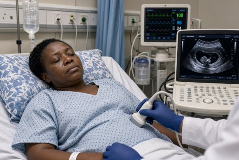
Ultrasound with Doppler assesses portal vein patency and flow direction, splenic size, and can suggest portal hypertension from an enlarged portal vein diameter and collateral vessels.
Upper GI endoscopy screens for esophageal and gastric varices at the time cirrhosis is diagnosed, with surveillance intervals guided by variceal size and liver disease severity.
Transient elastography and platelet count are used together in some algorithms (the Baveno criteria) to identify patients who can safely avoid screening endoscopy given a low probability of high risk varices.
Hepatic venous pressure gradient measurement, an invasive catheter based technique, provides the definitive hemodynamic assessment and is used in specialist settings, particularly for research or complex cases.
CT or MR imaging assesses vascular anatomy in suspected Budd-Chiari syndrome or portal vein thrombosis.
Differentials for the underlying cause include cirrhosis of any etiology, portal vein thrombosis (malignant or bland, often in a hypercoagulable state or with underlying cirrhosis or malignancy), schistosomiasis (periportal fibrosis, presinusoidal, preserved synthetic function relative to the degree of portal hypertension), and Budd-Chiari syndrome.