








Posterior Urethral Valves
Posterior urethral valves are congenital obstructing mucosal folds within the posterior urethra occurring exclusively in boys.
They are the commonest severe congenital cause of lower urinary tract obstruction in male infants.
Obstruction causes a spectrum of damage involving:
- Bladder hypertrophy
- Poor bladder compliance
- Vesicoureteric reflux
- Hydroureter
- Hydronephrosis
- Renal dysplasia
- Chronic kidney disease
Severe antenatal obstruction can cause oligohydramnios and secondary pulmonary hypoplasia.
Relief of the valve does not reverse congenital renal dysplasia. Long term renal and bladder surveillance is therefore required even after technically successful ablation.
Antenatal
Ultrasound may demonstrate:
- Bilateral hydroureteronephrosis
- Thick walled distended bladder
- Dilated posterior urethra
- Oligohydramnios
Neonatal
Presentation includes:
- Poor urinary stream
- Straining during urination
- Palpable bladder
- Respiratory failure from pulmonary hypoplasia
- Renal impairment
- Electrolyte disturbance
- Urinary tract infection or urosepsis
Older children can present with:
- Recurrent UTI
- Daytime urinary symptoms
- Enuresis
- Weak stream
- Chronic kidney disease
Initial laboratory assessment
Check:
- Serum creatinine
- Urea
- Sodium
- Potassium
- Bicarbonate
- Urinalysis
- Urine culture when infection is possible
Creatinine during the first days of life partially reflects maternal creatinine, so serial measurements are more informative than the first value alone.
The nadir creatinine after decompression is one of the strongest predictors of future renal outcome.
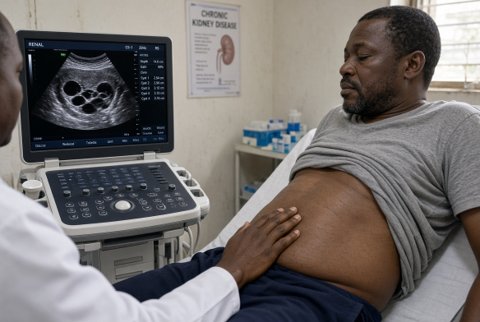
Ultrasound
Assess:
- Renal echogenicity
- Corticomedullary differentiation
- Hydronephrosis
- Ureteric dilatation
- Bladder wall
- Residual urine
Voiding cystourethrogram
VCUG establishes the diagnosis and can demonstrate:
- Dilated posterior urethra
- Abrupt narrowing at valve level
- Thick irregular bladder
- Vesicoureteric reflux
It also provides a baseline for future bladder management.
Immediate bladder drainage
If valves are suspected in a sick newborn, decompress the bladder promptly with a small urethral catheter.
Do not wait for definitive endoscopy before relieving severe obstruction.
Measure urine output after decompression.
A brisk post obstructive diuresis can produce major fluid and electrolyte losses.
Replace fluids carefully according to:
- Urine output
- Weight
- Sodium
- Potassium
- Acid base status
Treat UTI or urosepsis aggressively.
Definitive valve ablation
In a stable newborn of sufficient size for safe instrumentation, perform early cystoscopy and endoscopic valve ablation, often within the first 24 to 48 hours after stabilisation.
The obstructing valve leaflets are incised under direct vision.
Avoid deep circumferential urethral injury because this can cause postoperative urethral stricture.
After ablation:
- Leave bladder drainage according to operative findings
- Monitor creatinine
- Monitor urine output
- Repeat ultrasound
Small premature infant
Very small neonates may not accommodate safe endoscopic equipment.
If adequate urethral drainage cannot be maintained, perform temporary vesicostomy.
The vesicostomy creates a low pressure cutaneous bladder outlet and protects upper urinary tract drainage until later valve ablation becomes technically feasible.
Upper urinary tract diversion is rarely required and should be reserved for selected severe infection or drainage failure.
Vesicoureteric reflux
Do not automatically reimplant refluxing ureters immediately after valve diagnosis.
Reflux may improve after:
- Valve ablation
- Reduction in bladder pressure
- Bladder maturation
Initial management focuses on creating a low pressure bladder.
Valve bladder dysfunction
Bladder dysfunction can persist or worsen despite successful valve ablation.
Patterns include:
- Small hypertonic poorly compliant bladder
- Overactive bladder
- Large poorly contractile bladder with high residual urine
Management can include:
- Timed voiding
- Double voiding
- Antimuscarinic therapy
- Alpha blockade in selected outlet dysfunction
- Clean intermittent catheterisation
- Overnight bladder drainage
- Bladder augmentation in severe refractory low compliance disease
Antimuscarinic therapy
In a child with detrusor overactivity or poor compliance after confirmation of adequate emptying, a practical option is:
Oxybutynin approximately 0.2 mg/kg orally two to three times daily, titrated to age, response and adverse effects.
Monitor residual urine because reducing bladder contractions in a poorly emptying bladder can worsen retention.
Chronic kidney disease
Renal deterioration can continue even with a functioning urinary outlet because renal dysplasia was established antenatally.
Monitor lifelong:
- Blood pressure
- Creatinine and estimated GFR
- Proteinuria
- Electrolytes
- Growth
- Bladder function
- Hydronephrosis
Progressive end stage kidney disease may require dialysis and renal transplantation.
Optimising bladder pressure and emptying is essential before transplantation because a hostile high pressure bladder can damage the transplanted kidney.
