








Altered level of consciousness
Also known as: Reduced consciousness
Altered level of consciousness is any reduction in arousal or awareness, ranging from drowsiness through stupor to coma. It follows bilateral hemispheric dysfunction or dysfunction of the brainstem arousal system, and needs simultaneous resuscitation, assessment and treatment of reversible causes.
Anatomy and physiology. Arousal depends on the ascending reticular activating system in the upper pons and midbrain, projecting through the thalami to both cerebral hemispheres. Awareness depends on cortical function. A unilateral hemispheric lesion does not reduce consciousness unless it causes compression of the other hemisphere or brainstem.
Mechanisms. Diffuse metabolic or toxic suppression of both hemispheres, bilateral structural lesions, mass lesions causing herniation, and primary brainstem lesions.
Classification. Drowsiness (rousable to voice), stupor (rousable only to pain), coma (not rousable). Severity is scored with the Glasgow Coma Scale (eye opening 1 to 4, verbal response 1 to 5, motor response 1 to 6; total 3 to 15) and simplified as AVPU (alert, voice, pain, unresponsive). A score of 8 or less indicates coma and loss of airway protection.
Temporal patterns. Seconds (arrhythmia, seizure, subarachnoid haemorrhage, brainstem stroke), hours (hypoglycaemia, toxins, sepsis), and days to weeks (tumour, infection, hepatic or renal failure). Transient loss of consciousness with full recovery has separate causes, which are listed with the other differentials and tested for in the diagnostic approach.
Resuscitation first. Airway, breathing, circulation, glucose and temperature before detailed history.
History from witnesses, paramedics and records. Onset and circumstances, preceding headache, vomiting, seizure, chest pain, palpitations, trauma or fall, fever, diabetes and insulin use, alcohol.
Also ask about drug and medicine access (empty packets, opioid patches, sedatives), psychiatric history, epilepsy, cancer, HIV, anticoagulation, liver or kidney disease, travel, recent surgery, and exposure to smoke, solvents, pesticides, or a faulty heater.
Examination
- Vital signs: temperature, blood pressure, pulse, respiratory rate and pattern, oxygen saturation.
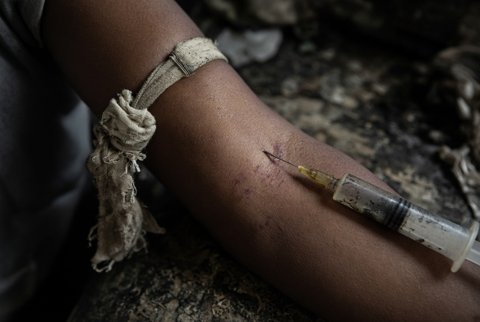
- Skin: needle marks, rash, petechiae, jaundice, cyanosis, burns, pressure sores, myxoedematous features, bruising or Battle sign and periorbital haematoma.
- Breath: ketones, alcohol, uraemic or hepatic fetor.
- Neurological: GCS, pupil size and reactivity, eye position, spontaneous and reflex eye movements (oculocephalic testing only after cervical spine injury is excluded), corneal and gag reflexes, facial symmetry, motor response and posturing, tone, reflexes, plantar responses, neck stiffness, fundi.
- Respiratory pattern: Cheyne Stokes breathing (bilateral hemispheric or metabolic), central neurogenic hyperventilation (midbrain), apneustic or ataxic breathing (pons or medulla).
- Pupils: pinpoint (opioids, pontine haemorrhage, organophosphates), unilateral fixed dilated (third nerve compression from uncal herniation), midposition fixed (midbrain), bilateral dilated (anticholinergics, sympathomimetics, severe hypoxia), reactive pupils with absent motor response favour a metabolic cause.
- Other: asterixis, tongue bite, signs of liver disease, thyroid, goitre scar, adrenal pigmentation, urinary retention.
Red flags
- GCS of 8 or less, or a fall of 2 or more points
- Anisocoria, fixed pupil, or abnormal eye position
- Decorticate or decerebrate posturing
- Irregular breathing, hypertension with bradycardia (Cushing response)
- Fever with neck stiffness or rash
- Seizures or non-convulsive status
- Hypotension, hypoxia, or hypoglycaemia
- Head or neck injury
- Suspected overdose or toxic exposure
- Pregnancy with hypertension
Special groups. Older adults: delirium superimposed on dementia and polypharmacy. Pregnancy: eclampsia, cerebral venous thrombosis, thrombotic thrombocytopenic purpura. Region specific: cerebral malaria, snakebite (neurotoxic and haemotoxic), organophosphate poisoning, methanol, carbon monoxide and African trypanosomiasis.
Structural, supratentorial
- Intracerebral haemorrhage, large hemispheric infarct with oedema, subarachnoid haemorrhage
- Subdural and extradural haematoma
- Tumour, abscess, hydrocephalus
- Cerebral venous sinus thrombosis, traumatic brain injury
Structural, infratentorial
- Basilar artery occlusion, pontine and cerebellar haemorrhage or infarct with brainstem compression
- Central pontine myelinolysis
Diffuse metabolic and endocrine
- Hypoglycaemia, diabetic ketoacidosis, hyperosmolar hyperglycaemic state
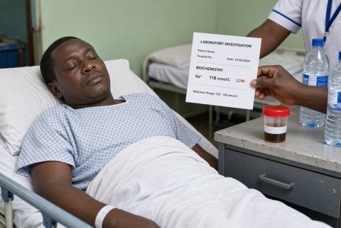
- Hyponatraemia, hypernatraemia, hypercalcaemia
- Uraemic encephalopathy, hepatic encephalopathy
- Hypoxia, hypercapnia
- Hypothermia, heat stroke
- Myxoedema coma, adrenal crisis, thyroid storm, Wernicke encephalopathy
Toxic and drug related
- Opioids, benzodiazepines, barbiturates, alcohol, gamma hydroxybutyrate
- Tricyclic antidepressants, antipsychotics, anticonvulsants, clonidine
- Paracetamol (late), salicylate, lithium
- Carbon monoxide, cyanide, organophosphates, methanol, ethylene glycol
Infective and inflammatory
- Bacterial meningitis, viral encephalitis, cerebral malaria, sepsis, brain abscess
- African trypanosomiasis, cryptococcal meningitis, tuberculous meningitis
- Autoimmune encephalitis, acute disseminated encephalomyelitis
Vascular and cardiac
- Cardiac arrest, high grade arrhythmia, shock of any cause
- Hypertensive encephalopathy, posterior reversible encephalopathy syndrome, eclampsia
Seizure related
- Post-ictal state, non-convulsive status epilepticus
Transient loss of consciousness
- Reflex, orthostatic and cardiac syncope; generalised seizure
- Basilar transient ischaemic attack, concussion, hypoglycaemia
Mimics
- Functional unresponsiveness, catatonia
- Locked-in syndrome, neuromuscular paralysis (Guillain Barré syndrome, myasthenic crisis, botulism) with preserved awareness
Reasoning. Treat life threats and give empirical therapy for hypoglycaemia, opioid toxicity and suspected CNS infection while testing. Then decide whether the cause is structural (focal signs, asymmetric motor or pupillary findings, posturing) or diffuse (symmetrical findings, preserved pupil reactions), and whether imaging should come before lumbar puncture.
Immediate bedside tests. Capillary glucose, oxygen saturation, temperature, ECG, blood pressure, urinary catheter or bladder scan, pregnancy test.
Blood tests.
Venous or arterial blood gas with lactate, co-oximetry for carboxyhaemoglobin and methaemoglobin, electrolytes, calcium, magnesium, phosphate, urea, creatinine, glucose, ketones, liver function, ammonia, full blood count, clotting, blood cultures, malaria test, creatine kinase and troponin.
Thyroid function and cortisol if endocrine cause is suspected. Ethanol, paracetamol and salicylate levels in any unexplained or suspected overdose.
Osmolality with calculation of the osmolar gap (over 10 mOsm/kg suggests toxic alcohols) and the anion gap (sodium minus chloride minus bicarbonate; over 12 mmol/L is raised).
Urine toxicology screens give limited, delayed information and do not change immediate care.
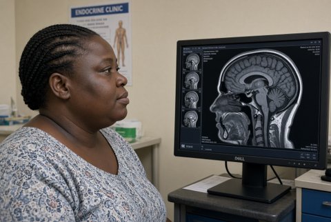
Imaging. Non-contrast CT head as soon as the patient is stable, when the cause is not clearly metabolic or toxic, with CT cervical spine after trauma. CT angiography or MRI with diffusion weighted imaging for suspected basilar occlusion (CT can be normal for hours). MRI for unexplained coma with normal CT (diffuse axonal injury, brainstem lesions, encephalitis, hypoxic injury).
Lumbar puncture after CT when meningitis or encephalitis is possible; give antibiotics first when CT or puncture would cause delay.
EEG for suspected non-convulsive status, which accounts for a minority of unexplained coma and responds to treatment.
Transient loss of consciousness. Eyewitness account, lying and standing blood pressure, glucose, 12 lead ECG for all; echocardiography when murmur, heart failure or abnormal ECG; ambulatory ECG monitoring; tilt testing when reflex syncope is suspected but unproven. High risk features: exertional or supine onset, palpitations before collapse, chest pain, family history of sudden death, abnormal ECG, structural heart disease, injury, and age over 65.
Limitations. Toxicology screening has false negatives. CT is insensitive early for posterior fossa infarction. A normal EEG does not exclude all non-convulsive status.
Airway and breathing. Intubation by a skilled operator when GCS is 8 or less, or when the airway cannot be protected, with cervical spine precautions after trauma. Oxygen to a saturation of 94 to 98 percent (88 to 92 percent when chronic hypercapnia is likely).
Circulation. IV access and fluid resuscitation for shock. Septic shock: crystalloid 30 mL/kg IV, broad spectrum antibiotics within 1 hour of recognition, and noradrenaline infusion for mean arterial pressure under 65 mmHg after fluids.
Empirical treatments given at once when the cause is unclear
- Glucose under 4 mmol/L: 10 percent glucose 150 to 200 mL IV over 15 minutes, repeated until over 4 mmol/L.
- Glucagon 1 mg IM when there is no IV access (ineffective in alcohol related, malnourished or liver disease hypoglycaemia).
- Give thiamine 500 mg IV every 8 hours for 3 days, before or with glucose, when alcohol misuse, malnutrition or vomiting is likely.
- Opioid toxicity (respiratory rate under 10 breaths/minute, pinpoint pupils): naloxone 0.4 mg IV, repeated every 2 to 3 minutes and titrated to respiratory rate of 10 or more, not to full arousal; IM 0.4 to 0.8 mg if no IV access.
- Use 0.04 to 0.1 mg doses in opioid dependent patients to avoid acute withdrawal. If effective, infuse two thirds of the effective bolus dose each hour.
- Observe for at least 4 to 6 hours after the last dose because naloxone acts for 30 to 90 minutes; longer for long acting opioids. If no response after 10 mg, reconsider the diagnosis.
- Flumazenil is not used in unknown overdose or in people who take benzodiazepines regularly because of seizure risk.
- Suspected meningitis or encephalitis: ceftriaxone 2 g IV every 12 hours, dexamethasone 10 mg IV every 6 hours for 4 days (first dose before or with the antibiotic).
- Add ampicillin 2 g IV every 4 hours if age over 60 or immunocompromised, and aciclovir 10 mg/kg IV every 8 hours for suspected encephalitis (adjust in renal impairment).
- Cerebral malaria: artesunate 2.4 mg/kg IV at 0, 12 and 24 hours, then once daily until oral therapy is possible, with glucose monitoring every 4 hours.
- Seizures: lorazepam 4 mg IV, repeated once after 10 minutes (maximum 8 mg), then levetiracetam 60 mg/kg IV (maximum 4.5 g) over 10 minutes if seizures continue.
- Adrenal crisis: hydrocortisone 100 mg IV immediately, then 50 mg IV every 6 hours, with 0.9 percent sodium chloride 1 L over the first hour.
- Diabetic ketoacidosis: 0.9 percent sodium chloride 1 L over the first hour, then fixed rate insulin 0.1 units/kg/hour once potassium is over 3.3 mmol/L, with potassium replacement to keep it at 4 to 5 mmol/L.
- Myxoedema coma: levothyroxine 200 to 400 micrograms IV as a loading dose (lower in older adults and in heart disease), then 1.6 micrograms/kg daily (75 percent of the dose IV), with hydrocortisone 100 mg IV every 8 hours until adrenal insufficiency is excluded.
- Carbon monoxide poisoning: 100 percent oxygen by non-rebreather mask until carboxyhaemoglobin is under 5 percent and symptoms resolve, and consider hyperbaric oxygen after loss of consciousness, neurological signs, carboxyhaemoglobin over 25 percent, or pregnancy.
- Organophosphate poisoning: atropine 1 to 2 mg IV, doubled every 5 minutes until secretions dry and the chest is clear, with decontamination.
- Paracetamol overdose needing treatment: acetylcysteine 150 mg/kg IV over 1 hour, then 50 mg/kg over 4 hours, then 100 mg/kg over 16 hours (cap weight at 110 kg).
- Hepatic encephalopathy: lactulose 30 to 45 mL orally 2 to 4 times daily titrated to 2 to 3 soft stools daily; treat the precipitant (bleeding, infection, electrolytes).
Raised intracranial pressure or herniation. Head of bed at 30 degrees, avoid hypotension and hypoxia, and arrange immediate neurosurgical referral.
- Mannitol 20 percent 0.5 to 1 g/kg IV over 15 to 20 minutes (stop if serum osmolality is over 320 mOsm/kg or in anuric renal failure).
- Or 3 percent sodium chloride 2 to 5 mL/kg IV over 10 to 20 minutes (monitor sodium, stop at over 155 to 160 mmol/L).
- Brief hyperventilation to a PaCO2 of 4.0 to 4.5 kPa as a bridge only.
Monitoring. GCS and pupils every 15 minutes initially, continuous ECG and oxygen saturation, glucose hourly until stable, sodium and osmolality during osmotherapy, temperature, urine output.
Referral and escalation. Intensive care for GCS 8 or less, haemodynamic instability or need for ventilation. Neurosurgery for mass lesions and hydrocephalus. Poisons advice and toxicology for overdose. Stroke team for suspected basilar occlusion.
Admission criteria. Every patient with an unexplained reduction in consciousness. Discharge only after transient loss of consciousness with a clear benign cause (typical reflex syncope with a normal ECG and examination) and safe follow up.
Safety netting. Return for any further collapse, confusion, weakness, speech or visual symptoms, severe headache or fever. Advise on driving after loss of consciousness according to local rules.
