








Fever with abdominal pain
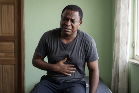
Fever with abdominal pain is a temperature of 38.0 degrees Celsius or over with acute or subacute abdominal pain, with or without vomiting, diarrhoea, jaundice or distension.
Classification by site and mechanism
- Hepatobiliary: acute cholecystitis, ascending cholangitis, liver abscess (pyogenic, amoebic), acute hepatitis, and perihepatitis (Fitz-Hugh-Curtis syndrome).
- Gastrointestinal: appendicitis, diverticulitis, infectious enterocolitis (Salmonella, Shigella, Campylobacter, Clostridioides difficile), typhoid enteric fever, inflammatory bowel disease, mesenteric adenitis, typhlitis, and perforated peptic ulcer.
- Peritoneal and retroperitoneal: peritonitis (primary, secondary, tertiary), spontaneous bacterial peritonitis, pancreatitis with necrosis, psoas abscess, and tuberculous peritonitis.
- Genitourinary and pelvic: pyelonephritis, pelvic inflammatory disease, tubo-ovarian abscess, prostatitis, and epididymo-orchitis.
- Vascular: mesenteric ischaemia, splenic infarction or abscess, and aortic infection.
- Thoracic and systemic: basal pneumonia, myocarditis, malaria, dengue, leptospirosis, sickle cell crisis, diabetic ketoacidosis, familial Mediterranean fever, and porphyria.
Epidemiology. Abdominal pain causes 5% to 10% of emergency visits, and fever raises the likelihood of infection or inflammation. Appendicitis has a lifetime risk of 7%, with a peak at 10 to 30 years. Cholecystitis and cholangitis occur in the setting of gallstones, which affect about 10% to 15% of adults. In older adults, abdominal pain has a higher rate of serious pathology (about 40% need surgery) with atypical features and muted fever.
Mechanisms. Visceral pain arises from distension or inflammation of hollow organs and is poorly localised (midline). Somatic parietal pain from peritoneal irritation is localised and worsened by movement. Referred pain follows shared nerve supply. Infection arises from obstruction (appendix, bile duct), mucosal breach (perforation, ischaemia), seeding from portal or systemic bacteraemia, or ascending infection from the genital tract.
Key distinctions. Pain migrating from the umbilicus to the right iliac fossa with anorexia suggests appendicitis. Right upper quadrant pain, fever and jaundice (Charcot triad) suggests cholangitis. Left iliac fossa pain with fever and altered bowel habit suggests diverticulitis. Fever, pain, ascites and encephalopathy in cirrhosis suggests spontaneous bacterial peritonitis.
Prolonged fever with abdominal pain, constipation then diarrhoea, relative bradycardia and rose spots suggests typhoid. Lower abdominal pain with cervical motion tenderness suggests pelvic inflammatory disease. Pain out of proportion to examination suggests mesenteric ischaemia.
History
- Pain: onset (sudden, gradual), site and migration, character (colic, constant, sharp), severity, radiation (to shoulder, back, groin), aggravating factors (movement, cough), relieving factors, and timing relative to meals.
- Associated symptoms: nausea, vomiting (bilious, faeculent), diarrhoea (bloody), constipation, absolute constipation and distension, anorexia, jaundice, dark urine, pale stool, weight loss, rigors, urinary symptoms, vaginal discharge, and haematemesis.
- Fever pattern: continuous, step ladder (typhoid), swinging (abscess), cyclical (malaria).
- Gynaecological history: last menstrual period, sexual history, contraception, pelvic pain, discharge, and pregnancy.
- Past history: gallstones, peptic ulcer, diverticular disease, hernia, abdominal surgery, inflammatory bowel disease, cirrhosis and ascites, sickle cell disease, HIV, diabetes, cancer, and tuberculosis.
- Drugs: nonsteroidal drugs, corticosteroids (mask signs), anticoagulants, antibiotics (Clostridioides difficile), opioids (constipation), immunosuppressants, and chemotherapy.
- Exposures and travel: contaminated food or water, sick contacts, freshwater exposure, raw milk, and tropical travel.
- Alcohol use.
Examination
- Vital signs: temperature, pulse, blood pressure, respiratory rate, oxygen saturation, mental state, urine output.
- General: dehydration, jaundice, pallor, lymph nodes, rose spots, stigmata of chronic liver disease, hernial orifices.
- Abdomen: inspection (distension, scars, visible peristalsis), palpation (guarding, rigidity, rebound, localised tenderness, Murphy sign, McBurney point, Rovsing sign), masses, hepatomegaly, splenomegaly, ascites, percussion, and bowel sounds (absent in ileus, high pitched in obstruction).
- Rectal examination (tenderness, mass, blood, melaena).
- Pelvic examination in women (cervical motion tenderness, adnexal mass, discharge).
- Genital and scrotal examination in men.
- Chest and cardiovascular examination (pneumonia, atrial fibrillation with embolism).
- Skin for petechiae, erythema nodosum and herpes zoster.
Red flags
- Hypotension, tachycardia, tachypnoea, confusion, mottled skin, oliguria, or lactate over 2 mmol/L (sepsis).
- Generalised peritonitis: board-like rigidity, rebound, silent abdomen, free air on imaging.
- Pain out of proportion to signs, especially in atrial fibrillation or vascular disease (mesenteric ischaemia).
- Jaundice with fever and rigors (cholangitis).
- Fever and abdominal pain in a patient with cirrhosis and ascites.
- Immunosuppression, neutropenia, HIV, steroids, or diabetes.
- Bilious vomiting, absolute constipation and distension (obstruction).
- Rectal bleeding, haematemesis, or melaena.
- Pregnancy or missed period with pain (ectopic pregnancy).
- Testicular pain (torsion) or pulsatile mass (aortic aneurysm).
- Age over 65 years with new pain.
- Postoperative fever with abdominal pain (anastomotic leak, abscess).
- Severe diarrhoea with distension and recent antibiotics (fulminant Clostridioides difficile colitis).
Special groups
- Children: appendicitis presents with vomiting, fever and diffuse pain; consider intussusception, mesenteric adenitis, and urinary infection.
- Older adults: blunted fever and signs, with higher rate of perforation and ischaemia.
- Pregnancy: appendix migrates upward. Consider cholecystitis, pyelonephritis, and ectopic pregnancy. Use ultrasound or magnetic resonance imaging.
- Immunosuppressed: typhlitis, cytomegalovirus colitis, and fungal infection, with minimal peritoneal signs.
- Cirrhosis: diagnostic paracentesis for every patient with ascites and fever or pain.
- Region specific: typhoid, amoebiasis, abdominal tuberculosis, schistosomiasis, hydatid disease, and cholera.
Hepatobiliary and pancreatic
- Acute calculous and acalculous cholecystitis.
- Ascending cholangitis and biliary sepsis.
- Pyogenic and amoebic liver abscess.
- Acute viral and alcoholic hepatitis.
- Fitz-Hugh-Curtis syndrome (perihepatitis) with pelvic inflammatory disease.
- Acute pancreatitis with infected necrosis or pseudocyst.
Gastrointestinal
- Appendicitis (perforated and appendiceal abscess).
- Diverticulitis with abscess or perforation.
- Perforated peptic ulcer.
- Infectious enterocolitis: Salmonella, Shigella, Campylobacter, Escherichia coli, Yersinia, Clostridioides difficile, cytomegalovirus, amoebiasis.
- Typhoid and paratyphoid fever.
- Inflammatory bowel disease (Crohn disease, ulcerative colitis with toxic megacolon).
- Intestinal obstruction with strangulation, mesenteric ischaemia, ischaemic colitis.
- Typhlitis in neutropenia.
- Intra-abdominal tuberculosis.
- Mesenteric adenitis.
Peritoneal and other
- Secondary bacterial peritonitis, spontaneous bacterial peritonitis, and tertiary peritonitis.
- Intra-abdominal and pelvic abscess; psoas abscess.
- Spontaneous or postoperative infection.
Genitourinary and gynaecological
- Pyelonephritis, infected renal stone, and pyonephrosis.
- Pelvic inflammatory disease and tubo-ovarian abscess.
- Ectopic pregnancy, ovarian torsion, and septic abortion.
- Prostatitis and epididymo-orchitis.
Systemic and metabolic
- Malaria, dengue, leptospirosis, and rickettsial disease.
- Sickle cell crisis and acute chest syndrome.
- Diabetic ketoacidosis.
- Familial Mediterranean fever, porphyria, and adrenal crisis.
- Lymphoma, hepatocellular carcinoma, and other malignancy with fever.
- Lower lobe pneumonia and myocardial infarction.
Step 1. Resuscitate and assess for surgical emergency. Establish intravenous access, give fluids, send blood cultures and lactate, and decide on analgesia and surgical review. Do not delay analgesia: opioids do not mask the diagnosis.
Step 2. First line tests
- Full blood count with differential, C-reactive protein, urea, creatinine, electrolytes, glucose, liver tests, amylase or lipase, calcium, lactate, coagulation, and group and save.
- Two sets of blood cultures before antibiotics.
- Urine dipstick and culture, and pregnancy test in women of reproductive age.
- Malaria rapid test or smear in endemic areas or recent travel.
- Erect chest radiograph (free subdiaphragmatic gas), and abdominal radiograph for suspected obstruction or toxic megacolon.
- Stool microscopy, culture and Clostridioides difficile toxin when diarrhoea is present.
- ECG in older adults and epigastric pain.
Step 3. Imaging
- Ultrasound as first imaging for right upper quadrant pain (gallstones, wall thickening, pericholecystic fluid, bile duct dilatation, liver abscess), pelvic pain, children and pregnancy.
- CT of the abdomen and pelvis with intravenous contrast for undifferentiated pain with fever in adults, suspected appendicitis (when ultrasound is inconclusive), diverticulitis, abscess, perforation, ischaemia, and pancreatitis.
- CT angiography for suspected mesenteric ischaemia.
- Magnetic resonance cholangiopancreatography for biliary obstruction with unclear cause.
- Magnetic resonance imaging in pregnancy when ultrasound is non diagnostic.
- Diagnostic paracentesis: ascitic fluid neutrophil count 250 cells/mm3 or over diagnoses spontaneous bacterial peritonitis, with culture in blood culture bottles at the bedside.
Step 4. Other tests
- Blood cultures and Widal are unreliable for typhoid; blood or bone marrow culture is the best test, and stool culture is positive in later weeks. Use typhoid polymerase chain reaction where available.
- Amoebic serology and stool antigen for amoebic liver abscess, with aspiration only for diagnostic doubt or failure to respond.
- Pelvic examination with endocervical swabs for chlamydia and gonorrhoea nucleic acid amplification, and transvaginal ultrasound for pelvic inflammatory disease.
- Endoscopy and colonoscopy (avoid in acute severe colitis with perforation risk) for inflammatory bowel disease.
- Tuberculosis testing: ascitic fluid adenosine deaminase, cytology, acid fast smear and culture, and peritoneal biopsy.
- Diagnostic laparoscopy when imaging is inconclusive and the patient worsens.
Step 5. Interpretation. White cell count and C-reactive protein support, but do not exclude, intra-abdominal infection. Normal tests do not exclude early appendicitis or ischaemia. Use serial examination and repeat imaging. A raised lactate suggests ischaemia or sepsis. Elevated lipase over 3 times the upper limit supports pancreatitis. Bilirubin, alkaline phosphatase and gamma-glutamyl transferase rise with cholangitis.
Limits. Fever and leucocytosis are absent in many older adults. Ultrasound depends on operator and body habitus. CT carries radiation and contrast risks in pregnancy and renal impairment. Blood cultures are positive in about a third of intra-abdominal sepsis.
Immediate
- Resuscitation: oxygen as needed, two large bore cannulae, and intravenous compound sodium lactate (Hartmann) solution 500 mL boluses, reassessing after each (30 mL/kg in septic shock or lactate 4 mmol/L or over). Keep nil by mouth. Insert a urinary catheter for urine output (target 0.5 mL/kg/hour). Add vasopressors (noradrenaline 0.05 to 0.5 micrograms/kg/minute) if mean arterial pressure remains under 65 mmHg after fluids.
- Analgesia: paracetamol 1 g intravenously every 6 hours (maximum 4 g daily; 3 g in patients under 50 kg), morphine 2.5 to 5 mg intravenously titrated every 5 to 10 minutes (reduce in older adults and renal impairment), and an antiemetic such as ondansetron 4 mg intravenously (check QTc if risk). Avoid nonsteroidal drugs in sepsis, acute kidney injury and bleeding risk.
- Antibiotics within 1 hour in sepsis and as soon as the diagnosis is made in others.
Empirical antibiotics for intra-abdominal infection (adjust to local resistance and results)
- Community acquired, mild to moderate: amoxicillin with clavulanic acid 1.2 g intravenously every 8 hours, or ceftriaxone 2 g once daily with metronidazole 500 mg intravenously every 8 hours. With penicillin allergy (nonsevere): ceftriaxone with metronidazole; severe allergy: ciprofloxacin 400 mg intravenously every 12 hours with metronidazole, or gentamicin 5 mg/kg once daily with metronidazole (specialist advice).
- Severe, healthcare associated or high risk: piperacillin with tazobactam 4.5 g intravenously every 6 hours (creatinine clearance 20 to 40 mL/min, 4.5 g every 8 hours; under 20 mL/min, 4.5 g every 12 hours), or meropenem 1 g every 8 hours (with renal dose reduction, and for extended spectrum beta-lactamase producers). Add vancomycin when Enterococcus faecium or MRSA is likely, and an antifungal (fluconazole 800 mg loading then 400 mg daily, or caspofungin 70 mg then 50 mg daily) for yeast in peritoneal fluid in critically ill patients.
- Duration: after adequate source control, 4 days (fixed short course) for most intra-abdominal infections; longer (7 to 14 days) for undrained abscess, ongoing signs, or immunosuppression.
Treatment by diagnosis
- Appendicitis: appendicectomy (laparoscopic) within 24 hours, with preoperative antibiotics (ceftriaxone with metronidazole, single dose) and continued therapy for perforation (3 to 5 days after surgery). Antibiotic only management is possible in selected uncomplicated cases with a risk of recurrence of about 30% in 1 year. Appendiceal abscess: percutaneous drainage and antibiotics, interval appendicectomy considered.
- Acute cholecystitis: antibiotics (amoxicillin with clavulanic acid, or ceftriaxone with metronidazole) and early laparoscopic cholecystectomy within 7 days of onset (or within 72 hours when possible). Percutaneous cholecystostomy in unfit patients.
- Ascending cholangitis: resuscitation, piperacillin with tazobactam 4.5 g every 6 hours (or ceftriaxone 2 g with metronidazole), and biliary drainage by endoscopic retrograde cholangiopancreatography within 24 hours for severe disease (within 48 to 72 hours in moderate disease). Percutaneous transhepatic drainage if endoscopy fails.
- Diverticulitis: uncomplicated, mild: oral amoxicillin with clavulanic acid 625 mg three times daily for 5 to 7 days (or observation without antibiotics in selected patients), clear fluids; complicated (abscess over 4 cm): percutaneous drainage and intravenous antibiotics; perforation with peritonitis: surgery (Hartmann procedure or primary anastomosis).
- Perforated viscus and generalised peritonitis: emergency laparotomy or laparoscopy, source control, peritoneal lavage, and intensive care.
- Spontaneous bacterial peritonitis: ceftriaxone 2 g intravenously once daily for 5 days (cefotaxime 2 g every 8 hours as alternative), with albumin 1.5 g/kg on day 1 and 1 g/kg on day 3. Secondary prophylaxis with norfloxacin 400 mg or ciprofloxacin 500 mg daily after an episode. Stop diuretics and nephrotoxic drugs during the episode.
- Typhoid fever: ceftriaxone 2 g intravenously once daily for 10 to 14 days, or azithromycin 1 g orally on day 1 then 500 mg daily for 6 days (uncomplicated disease), with higher risk of resistance to ciprofloxacin and chloramphenicol in many regions. Add dexamethasone 3 mg/kg then 1 mg/kg every 6 hours for 8 doses for shock or encephalopathy. Surgical review for intestinal perforation (usually in the second to third week).
- Amoebic liver abscess: metronidazole 800 mg orally three times daily (or 500 mg intravenously every 8 hours) for 7 to 10 days, followed by paromomycin 500 mg three times daily for 7 days (or diloxanide furoate 500 mg three times daily for 10 days) to clear luminal cysts. Aspirate when the abscess is over 5 cm, in the left lobe, or if there is no response in 72 hours. Pyogenic abscess: percutaneous drainage and 4 to 6 weeks of antibiotics.
- Pelvic inflammatory disease: ceftriaxone 1 g intramuscularly or intravenously once daily, plus doxycycline 100 mg orally twice daily and metronidazole 400 mg orally twice daily for 14 days. Admit when severe, pregnant, unable to take oral therapy, or tubo-ovarian abscess is present. Treat partners and test for HIV and syphilis. Fitz-Hugh-Curtis syndrome is treated with the same regimen.
- Infectious enterocolitis: oral or intravenous rehydration; antibiotics only for severe or invasive disease, immunosuppressed, or with sepsis (azithromycin 500 mg daily for 3 days, or ciprofloxacin 500 mg twice daily for 3 days if susceptible). Clostridioides difficile: oral vancomycin 125 mg four times daily for 10 days, or fidaxomicin 200 mg twice daily for 10 days; fulminant disease: vancomycin 500 mg orally four times daily with metronidazole 500 mg intravenously every 8 hours, and surgical review.
- Abdominal tuberculosis: isoniazid 5 mg/kg (maximum 300 mg), rifampicin 10 mg/kg (maximum 600 mg), pyrazinamide 25 mg/kg and ethambutol 15 mg/kg daily for 2 months, then isoniazid and rifampicin for 4 months (up to 9 months in some cases), with pyridoxine.
- Mesenteric ischaemia: resuscitation, broad spectrum antibiotics, heparin infusion, and vascular surgical or radiological revascularisation.
- Malaria with abdominal pain: artesunate 2.4 mg/kg intravenously at 0, 12 and 24 hours then daily for severe disease, or artemether with lumefantrine for uncomplicated disease.
- Ectopic pregnancy, ovarian torsion, and testicular torsion: urgent surgical or gynaecological management.
Special populations
- Pregnancy: ceftriaxone, amoxicillin with clavulanic acid or cefuroxime for infection; avoid fluoroquinolones, tetracyclines and aminoglycosides unless essential; surgery should not be delayed because of pregnancy.
- Children: appendicitis treated with surgery and ceftriaxone 50 mg/kg daily (maximum 2 g) with metronidazole 7.5 mg/kg every 8 hours.
- Older adults: lower threshold for CT, early surgical review, and renal dose adjustment.
- Immunosuppressed: broaden antimicrobial cover, early imaging, and fungal and viral testing.
- Cirrhosis and renal impairment: avoid nephrotoxins, adjust doses, and give albumin.
Monitoring. Vital signs hourly in sepsis, urine output, lactate repeated within 2 to 4 hours, serial abdominal examination every 2 to 4 hours, white cell count and C-reactive protein daily, creatinine, culture results, drain output, and imaging if no improvement in 48 to 72 hours.
Referral and escalation. General surgery for peritonitis, appendicitis, perforation, obstruction and abscess. Gastroenterology for cholangitis, inflammatory bowel disease and complicated colitis. Interventional radiology for drainage. Infectious diseases for resistant organisms, immunosuppression, typhoid, amoebiasis and tuberculosis. Gynaecology for pelvic causes. Intensive care for organ dysfunction.
Admission criteria. Sepsis, peritonitis, suspected surgical diagnosis, jaundice with fever, inability to take oral intake, immunosuppression, cirrhosis with fever, pregnancy, elderly patients with new pain and fever, and failure of outpatient management.
Discharge and follow up. Discharge only when afebrile, pain controlled, eating and drinking, and a diagnosis or safe follow up plan is in place. Review at 24 to 48 hours. Complete oral antibiotics. Safety net advice: return for worsening pain, persistent vomiting, rigors, jaundice, blood in stool, fainting, or distension.
