










Raised jugular venous pressure
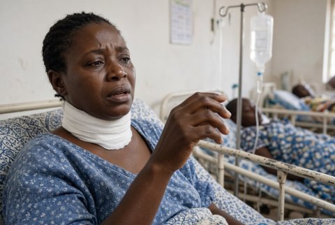
The jugular venous pressure reflects right atrial (central venous) pressure. It is raised when the top of the internal jugular venous column is more than 3 cm vertically over the sternal angle with the patient semi recumbent at 45 degrees. It is a sign of volume overload, right heart failure, pericardial disease, obstruction to right heart filling or outflow, or superior vena cava obstruction.
In equivalent terms, a raised pressure is a mean right atrial pressure over 8 mmHg, or the column visible over the clavicle when sitting at 90 degrees.
Clinical relevance. A raised jugular venous pressure is among the most reliable bedside signs of elevated filling pressures. It predicts admission and death in heart failure.
Some causes are immediately life threatening and treatable: cardiac tamponade, massive pulmonary embolism, tension pneumothorax and right ventricular infarction. Waveform analysis gives specific clues (cannon a waves, giant v waves, steep y descent).
Classification by mechanism.
- Raised right ventricular filling pressure from impaired right ventricular function: right heart failure (left heart failure, pulmonary hypertension, cor pulmonale, right ventricular infarction, cardiomyopathy), tricuspid valve disease (regurgitation, stenosis), pulmonary valve stenosis.
- Restricted filling: cardiac tamponade, constrictive pericarditis, restrictive cardiomyopathy, tension pneumothorax, severe asthma and chronic obstructive pulmonary disease with air trapping, mechanical ventilation with high pressure, massive pleural effusion.
- Obstruction: massive pulmonary embolism, superior vena cava obstruction (the pressure is raised in the neck and upper body veins without pulsation), right atrial tumour or thrombus, mediastinal mass.
- Volume overload: fluid overload (renal failure, nephrotic syndrome, iatrogenic fluid), high output states (arteriovenous fistula, thyrotoxicosis, anaemia, Paget disease, beriberi).
- Rhythm related: atrioventricular dissociation, atrial fibrillation (absent a wave), complete heart block (cannon a waves), junctional rhythm.
Waveform components. The a wave (atrial contraction), c wave (tricuspid closure), x descent (atrial relaxation), v wave (atrial filling), y descent (tricuspid opening and atrial emptying). The normal jugular venous pressure falls on inspiration; paradoxical rise on inspiration is Kussmaul sign.
History.
- Breathlessness, orthopnoea, paroxysmal nocturnal dyspnoea, ankle and abdominal swelling, weight gain, fatigue, early satiety, right upper quadrant pain, nausea, nocturia, palpitations, chest pain, syncope.
- Features of tamponade: pleuritic chest pain, fever, breathlessness, recent myocardial infarction, trauma, cardiac surgery, cancer, tuberculosis, renal failure, anticoagulation, aortic dissection.
- Features of pulmonary embolism: sudden breathlessness, pleuritic pain, haemoptysis, leg swelling, immobilisation, cancer, previous thrombosis.
- Chronic lung disease, smoking, sleep apnoea, previous tuberculosis, asbestos exposure, chest radiotherapy, cardiac surgery.
- Facial swelling, headache and neck fullness worse on bending or lying (superior vena cava obstruction), cough, weight loss, dysphagia, central line or pacemaker lead, haemoptysis.
- Drugs and fluids: intravenous fluids, NSAIDs, pioglitazone, calcium channel blockers, corticosteroids, anthracyclines, trastuzumab.
- Past history: heart failure, valve disease, kidney and liver disease, thyroid disease, rheumatic fever, connective tissue disease.
Examination.
- Method: place the patient semi recumbent at 45 degrees (adjust the angle until the venous pulsation is visible), with the head turned slightly to the left and good lighting.
- Examine the right internal jugular vein (medial to the sternocleidomastoid). It has a double waveform, is obliterated by pressure at the base, varies with respiration and posture, and is not palpable.
- Measure the vertical height of the top of the pulsation over the sternal angle. Add 5 cm to estimate right atrial pressure in cmH2O.
- Hepatojugular (abdominojugular) reflux: firm pressure on the right upper abdomen for 10 seconds produces a sustained rise of over 3 cm in the neck vein in right heart failure.
- Kussmaul sign (rise on inspiration), Pemberton sign (facial plethora and venous distension when arms are raised over the head; superior vena cava obstruction).
- Giant v waves: tricuspid regurgitation, with pulsatile liver and ear lobe movement.
- Cannon a waves: atrioventricular dissociation, ventricular tachycardia, pacing, complete heart block, junctional rhythm.
- Absent a wave: atrial fibrillation. Prominent a wave: pulmonary hypertension, pulmonary stenosis, tricuspid stenosis.
- Steep x and y descents: constrictive pericarditis (Friedreich sign). Slow y descent: tricuspid stenosis. Tamponade shows absent y descent.
- Cardiac: heart rate and rhythm, blood pressure, pulsus paradoxus (fall of over 10 mmHg in systolic pressure on inspiration), muffled heart sounds, rub, third heart sound, loud P2, right ventricular heave, murmurs (tricuspid regurgitation, pulmonary stenosis, mitral stenosis), pericardial knock.
- Chest: crackles, wheeze, effusion, hyperinflation, absent breath sounds, tracheal deviation.
- Abdomen: hepatomegaly (pulsatile in tricuspid regurgitation), ascites, splenomegaly; peripheral oedema, sacral oedema, cool peripheries.
- Face and neck: swollen face, dilated chest wall veins (superior vena cava obstruction, venous collaterals flow downward), thyroid, lymph nodes.
- Signs of cause: clubbing, cyanosis, cachexia, jaundice, stigmata of endocarditis.
Red flags.
- Hypotension with raised jugular venous pressure and muffled heart sounds (Beck triad; tamponade), pulsus paradoxus, tachycardia, breathlessness.
- Sudden breathlessness, hypoxia, pleuritic pain, tachycardia, or syncope (massive pulmonary embolism).
- Hypotension, hyperresonance, absent breath sounds and tracheal deviation (tension pneumothorax).
- Inferior myocardial infarction with raised jugular venous pressure, clear lungs and hypotension (right ventricular infarction).
- Stridor, facial swelling and plethora (superior vena cava obstruction with airway or cerebral oedema).
- Kussmaul sign with calcified pericardium or past tuberculosis (constriction).
- Giant v waves with new murmur and fever (endocarditis).
- Haemodynamic instability, oliguria, confusion.
Cardiac.
- Right heart failure from left heart failure (commonest).
- Cor pulmonale and pulmonary hypertension (chronic lung disease, pulmonary embolism, sleep apnoea, idiopathic pulmonary arterial hypertension, left heart disease, chronic thromboembolic disease).
- Right ventricular infarction.
- Cardiomyopathy (dilated, restrictive, arrhythmogenic).
- Tricuspid regurgitation and stenosis, pulmonary stenosis.
- Atrial myxoma, Ebstein anomaly, Eisenmenger syndrome, atrial septal defect with pulmonary hypertension, myocarditis.
Pericardial.
- Cardiac tamponade (effusion from malignancy, tuberculosis, uraemia, haemorrhage, dissection, post cardiac surgery, trauma, infection, connective tissue disease).
- Constrictive pericarditis (tuberculosis, radiation, surgery, idiopathic).
- Effusive constrictive pericarditis.
Obstructive and thoracic.
- Massive pulmonary embolism, tension pneumothorax.
- Severe asthma or chronic obstructive pulmonary disease with dynamic hyperinflation.
- Large pleural effusion.
- High intrathoracic pressure (mechanical ventilation, positive end expiratory pressure).
- Mediastinal mass.
- Superior vena cava obstruction (lung cancer, lymphoma, central venous catheter thrombosis, fibrosing mediastinitis, goitre).
- Right atrial thrombus.
Volume and high output. Fluid overload (renal failure, nephrotic syndrome, intravenous fluids, transfusion), hepatic cirrhosis with hepatorenal syndrome, arteriovenous fistula, thyrotoxicosis, anaemia, beriberi, pregnancy, Paget disease.
Rhythm. Complete heart block, ventricular tachycardia with atrioventricular dissociation, junctional rhythm, pacing (cannon a waves).
Mimics. Short or thick neck, obesity, wrong patient position, carotid pulsation mistaken for jugular venous pressure, external jugular vein distension from head down position.
Clue | Think of |
|---|---|
Raised jugular venous pressure, oedema, third heart sound, crackles | Congestive heart failure |
Raised pressure, hypotension, muffled sounds, pulsus paradoxus, absent y descent | Cardiac tamponade |
Raised pressure rising on inspiration (Kussmaul), steep y descent, pericardial knock | Constrictive pericarditis |
Sudden breathlessness, pleuritic pain, clear lungs, raised pressure | Massive pulmonary embolism |
Inferior infarct, hypotension, clear lungs, raised pressure | Right ventricular infarction |
Giant v waves, pulsatile liver, pansystolic murmur | Tricuspid regurgitation |
Cannon a waves, irregular intermittent | Atrioventricular dissociation |
Absent a wave, irregularly irregular pulse | Atrial fibrillation |
Non pulsatile raised pressure, facial plethora, Pemberton sign | Superior vena cava obstruction |
Hyperinflation, wheeze, smoker, loud P2 | Cor pulmonale |
Raised pressure, hypotension, tracheal deviation, absent breath sounds | Tension pneumothorax |
Raised pressure, oedema, proteinuria, oliguria | Fluid overload (renal) |
- Treat unstable patients first; assess airway, breathing, circulation, saturation, blood pressure and perfusion, then examine the neck veins.
- Repeat the jugular venous examination at the correct angle; confirm that the pulsation is venous (double waveform, obliterated by pressure, changes with posture and respiration).
- Do a 12 lead ECG. Look for right ventricular strain pattern, S1Q3T3, right bundle branch block, low voltage and electrical alternans in effusion, ST elevation inferior infarct with right sided leads V3R and V4R, conduction block, and atrial fibrillation.
- Do a chest radiograph. Look for cardiomegaly, pulmonary congestion, hyperinflation, pneumothorax, effusion, mediastinal widening or mass, and calcified pericardium.
- Bedside echocardiography in all with raised jugular venous pressure and instability. Assess pericardial effusion and right atrial or ventricular diastolic collapse, and right ventricular size and function (McConnell sign, septal flattening in embolism).
- Echocardiography also assesses tricuspid regurgitation and pulmonary pressure, left ventricular function, valve disease, and intracardiac thrombus or mass.
- Inferior vena cava size and collapse: plethoric, non collapsing over 2.1 cm suggests right atrial pressure 15 mmHg.
- Blood tests: full blood count, urea, creatinine, electrolytes, liver function, albumin, natriuretic peptide, troponin, D-dimer when pulmonary embolism is possible, thyroid stimulating hormone, glucose, lactate, blood gas, coagulation.
- CT pulmonary angiography (pulmonary embolism, chronic thromboembolic disease), contrast CT chest (superior vena cava obstruction, mediastinal mass, pericardial thickening and calcification), cardiac MRI (constriction, cardiomyopathy, right ventricular dysplasia, infiltration).
- Pericardiocentesis for tamponade with fluid analysis (cell count, protein, lactate dehydrogenase, cytology, culture and mycobacterial testing, adenosine deaminase over 40 units/L in tuberculosis).
- Right heart catheterisation to define pressures and differentiate constriction from restriction (dip and plateau, interdependence), pulmonary hypertension and shunts.
- Pulmonary function tests, sleep studies, V/Q scan, autoimmune and HIV tests, as the cause demands.
- Venography for suspected superior vena cava obstruction when needed before stenting.
Limits. Jugular venous pressure is hard to judge in obesity, short necks and severe tricuspid regurgitation (pulsation is higher than expected). It is also hard to judge in patients with a very high pressure, where the pulsation may be up to the jaw and the patient needs to sit upright.
Inspiratory fall may be hard to appreciate. A normal pressure does not exclude heart failure after diuretics or in orthopnoeic compensation.
Cardiac tamponade (emergency).
- Sit up, oxygen only if hypoxic, intravenous access. Avoid positive pressure ventilation and diuretics. Give small boluses of 0.9% sodium chloride 250 to 500 mL if hypotensive (to maintain preload).
- Urgent pericardiocentesis under echocardiography (subxiphoid or apical), with the drain left in situ until output is under 25 to 50 mL/day.
- Surgical drainage (pericardial window) for haemorrhagic, traumatic, post cardiac surgery or recurrent effusion.
- Aortic dissection with tamponade needs emergency surgery. Avoid pericardiocentesis delaying repair, or limit drainage.
- Treat the cause.
- Tuberculosis: isoniazid 5 mg/kg to a maximum of 300 mg, rifampicin 10 mg/kg to a maximum of 600 mg, pyrazinamide 25 mg/kg to a maximum of 2 g, ethambutol 15 mg/kg for 2 months, then isoniazid and rifampicin for 4 months. Add pyridoxine 10 to 25 mg daily, and prednisolone 1 mg/kg daily for 4 weeks then tapering over 8 weeks.
- Uraemia: dialysis. Malignancy: oncology, sclerosis. Bacterial pericarditis: drainage and antibiotics.
- Idiopathic or viral pericarditis: ibuprofen 600 mg three times daily for 1 to 2 weeks with colchicine 500 micrograms twice daily (500 micrograms daily if under 70 kg) for 3 months, and omeprazole 20 mg daily.
Massive pulmonary embolism with shock.
- Oxygen, cautious fluids (up to 500 mL), noradrenaline 0.05 to 0.5 micrograms/kg/minute for hypotension.
- Unfractionated heparin 80 units/kg bolus then 18 units/kg/hour.
- Systemic thrombolysis with alteplase 100 mg over 2 hours (10 mg bolus then 90 mg over 2 hours; or 0.6 mg/kg over 15 minutes, maximum 50 mg, in cardiac arrest) unless contraindicated.
- Catheter directed therapy or surgical embolectomy when thrombolysis is contraindicated or fails.
- Then oral anticoagulation: rivaroxaban 15 mg twice daily for 21 days then 20 mg daily; or apixaban 10 mg twice daily for 7 days then 5 mg twice daily.
Tension pneumothorax. Immediate needle decompression (large bore cannula, second intercostal space mid clavicular line or fifth intercostal space anterior axillary line), then a chest drain.
Right ventricular infarction.
- Avoid nitrates, diuretics and opioids that lower preload.
- Give 0.9% sodium chloride 250 to 500 mL boluses (up to 1 to 2 litres) with reassessment.
- Dobutamine 2.5 to 10 micrograms/kg/minute if hypotension persists.
- Maintain atrioventricular synchrony (pacing for heart block, cardioversion for atrial fibrillation).
- Urgent reperfusion by percutaneous coronary intervention.
Superior vena cava obstruction.
- Sit upright, oxygen.
- Dexamethasone 8 to 16 mg intravenously (for lymphoma or steroid sensitive tumours; avoid before biopsy if possible).
- Endovascular stenting (rapid relief).
- Radiotherapy or chemotherapy for the underlying cancer (small cell lung cancer, lymphoma).
- Anticoagulation for thrombosis, catheter removal or thrombolysis.
- Airway or cerebral oedema is an emergency.
Acute or decompensated heart failure and fluid overload.
- Furosemide 40 mg intravenously (20 to 40 mg if diuretic naive; at least the usual oral dose intravenously for chronic users, often 40 to 80 mg), repeated or infusion 5 to 40 mg/hour, with metolazone 2.5 to 5 mg.
- Glyceryl trinitrate infusion 10 to 200 micrograms/minute if systolic pressure over 110 mmHg.
- Non invasive ventilation for respiratory distress. Renal replacement or ultrafiltration for refractory overload.
- Sodium restriction under 2 g/day and fluid restriction 1.5 to 2 litres in refractory disease. Daily weights.
Long term heart failure with reduced ejection fraction.
- ACE inhibitor (ramipril 2.5 mg twice daily increasing to 5 mg twice daily) or sacubitril-valsartan 24/26 mg twice daily increasing to 97/103 mg twice daily.
- Beta blocker (bisoprolol 1.25 mg daily increasing to 10 mg).
- Spironolactone 25 mg daily.
- Dapagliflozin 10 mg or empagliflozin 10 mg daily.
Right heart failure from cor pulmonale and pulmonary hypertension.
- Treat hypoxia (long term oxygen when saturation is 88% or less).
- Diuretics (furosemide 40 to 80 mg daily with spironolactone 25 to 100 mg). Avoid excessive diuresis.
- Treat the lung disease or sleep apnoea.
- Anticoagulation for chronic thromboembolic disease, and pulmonary artery balloon angioplasty or endarterectomy.
- Specialist drugs for pulmonary arterial hypertension after right heart catheterisation: sildenafil 20 mg three times daily, bosentan 62.5 mg twice daily then 125 mg twice daily, macitentan 10 mg daily.
Tricuspid regurgitation. Diuretics, treat the cause (endocarditis: antibiotics; pulmonary hypertension; left heart disease), surgery or transcatheter repair for severe symptomatic disease.
Constrictive pericarditis and restrictive cardiomyopathy.
- Diuretics for congestion (furosemide 40 to 80 mg daily).
- Pericardiectomy for symptomatic constriction after confirming the diagnosis.
- Tuberculous disease needs the antituberculous regimen before and after surgery: isoniazid 5 mg/kg to a maximum of 300 mg, rifampicin 10 mg/kg to a maximum of 600 mg, pyrazinamide 25 mg/kg to a maximum of 2 g and ethambutol 15 mg/kg for 2 months, then isoniazid and rifampicin for 4 months, with pyridoxine.
- Restrictive cardiomyopathy: treat the cause and diurese cautiously. Amyloid: tafamidis 61 mg daily for transthyretin; chemotherapy for light chain disease. Iron chelation for haemochromatosis; steroids for sarcoidosis.
Rhythm disturbances.
- Atropine 500 micrograms intravenously (repeat to a maximum of 3 mg) or pacing for symptomatic bradycardia and complete heart block.
- Amiodarone 300 mg intravenously then 900 mg over 24 hours for ventricular tachycardia.
- Cardioversion for unstable tachyarrhythmia.
- Anticoagulation and rate or rhythm control for atrial fibrillation.
Renal and hepatic causes. Dialysis or ultrafiltration. Treat nephrotic syndrome (loop diuretic, albumin, specific therapy). For cirrhosis with ascites use spironolactone 100 mg with furosemide 40 mg, and paracentesis with albumin 8 g for each litre removed over 5 litres.
Monitoring. Continuous ECG, blood pressure, oxygen saturation and urine output in unstable patients. Daily weight, fluid balance, jugular venous pressure and peripheral oedema. Urea, creatinine and electrolytes daily during diuresis, then 1 to 2 weeks after changes. Repeat echocardiography after pericardiocentesis (24 hours) and at 1 to 3 months.
Referral and escalation. Cardiology urgently for tamponade, constriction, right ventricular infarction, new heart failure and tricuspid disease. Respiratory and pulmonary hypertension centres for pulmonary vascular disease. Cardiothoracic surgery for effusion drainage and pericardiectomy. Oncology for malignant effusion and superior vena cava obstruction. Nephrology for renal overload. Critical care for shock.
Admission. All patients with raised jugular venous pressure and instability, tamponade, suspected pulmonary embolism, right ventricular infarction, superior vena cava obstruction, new or decompensated heart failure, severe fluid overload, arrhythmia or syncope.
Safety netting. Return at once for breathlessness at rest, chest pain, fainting, a rapid heartbeat, facial or arm swelling, severe headache, fever, reduced urine, or weight gain over 2 kg in 3 days. Take diuretics as prescribed, weigh daily, and limit salt.
Special groups.
- Pregnancy: peripartum cardiomyopathy, pulmonary embolism, aortic dissection and Eisenmenger syndrome. Avoid ACE inhibitors, angiotensin receptor blockers and spironolactone (use furosemide, beta blocker, hydralazine with nitrate). Anticoagulate with enoxaparin 1 mg/kg twice daily.
- Children: congenital heart disease, myocarditis and pericardial effusion.
- Older adults: heart failure with preserved ejection fraction, amyloidosis and renal disease; observe fluid balance carefully.
- Intensive care patients: raised pressure from positive pressure ventilation and fluid load.
- Endemic regions: tuberculous pericardial disease, rheumatic valve disease, endomyocardial fibrosis, Chagas cardiomyopathy and schistosomal pulmonary hypertension are common causes.
