









Salivary gland swelling
Also known as: Parotid swelling, Submandibular swelling
Salivary gland swelling is enlargement of the parotid, submandibular, sublingual or minor salivary glands. It may be acute and painful (infection, obstruction), recurrent (stone, duct stricture), chronic (autoimmune, systemic) or a mass.
Anatomy and physiology. Parotid glands (serous) lie in front of the ear and drain via Stensen duct opposite the upper second molar; they are traversed by the facial nerve branches. The submandibular glands (mixed) drain via Wharton duct on the floor of the mouth, and have the highest rate of stones (80 to 90 percent of sialoliths). Sublingual and about 600 to 1,000 minor glands are mucous.
Classification. Acute sialadenitis (viral, bacterial), obstructive (stones, strictures), chronic sialadenitis and recurrent parotitis, autoimmune (Sjogren syndrome, IgG4 related disease), systemic and metabolic (sialosis), granulomatous and infiltrative, benign tumours, malignant tumours, and non-salivary masses (lymph nodes, cysts, lipoma).
Key distinctions. Pain and swelling with meals indicates obstruction. Fever, erythema and pus from the duct indicate acute bacterial sialadenitis. Bilateral painful swelling with mumps contact indicates viral parotitis. A firm painless mass, facial weakness, or skin fixation indicates malignancy.
History. Ask about:
- Onset, duration, side (unilateral, bilateral), pain, and relationship to eating (colic).
- Fever, bad taste, dry mouth and eyes, joint pain, rash, facial weakness, trismus and dysphagia.
- Mass growth, weight loss, previous radiotherapy (including radioiodine) and skin cancer.
- Mumps exposure and vaccination, HIV risk, alcohol, eating disorders (bulimia), diabetes, dehydration, and drug history.
Drug history. Anticholinergics, antihistamines, antidepressants, diuretics, antihypertensives, and drugs causing parotid swelling (iodine contrast, thiouracil, phenothiazines, isoprenaline, phenylbutazone); radioiodine.
Examination. Inspect and palpate the glands bimanually (intraoral and extraoral). Assess:
- Duct orifices (pus, erythema, stone, milking gland).
- Size, consistency (firm, rubbery, hard, cystic), fixity, tenderness and skin.
- Facial nerve function (all branches) and cervical nodes.
- Oral dryness, dentition, eyes (Sjogren), joints and skin.
- Trismus and parapharyngeal bulge.
Red flags
- Facial nerve weakness, pain with numbness, or skin fixation
- Rapidly enlarging, hard, fixed or painless mass
- Cervical lymphadenopathy with a gland mass
- Airway threat, floor of mouth swelling with fever (Ludwig angina)
- Fever, rigors and sepsis with tender gland
- History of skin cancer in the drainage area, or previous radiotherapy
- Bilateral swelling with systemic symptoms (lymphoma, sarcoid, HIV)
- Children with a persistent unilateral mass
Special groups. Children: mumps, juvenile recurrent parotitis, HIV, haemangioma, lymphatic malformation. Pregnancy: sialosis. Older adults: dehydration related parotitis after surgery, and malignancy. Region specific: mumps where vaccination is incomplete, HIV related salivary gland disease and tuberculosis.
Infective and inflammatory
- Mumps and other viral parotitis (Epstein Barr virus, cytomegalovirus, coxsackievirus, parainfluenza, influenza, HIV)
- Acute bacterial sialadenitis (Staphylococcus aureus, streptococci, anaerobes), acute suppurative parotitis, parotid abscess
- Chronic sialadenitis, recurrent parotitis (children and adults)
- Tuberculosis, actinomycosis, atypical mycobacteria, cat scratch disease, syphilis, toxoplasmosis
- HIV related lymphoepithelial cysts and diffuse infiltrative lymphocytosis syndrome
Obstructive
- Sialolithiasis (submandibular, parotid), salivary duct stricture, mucus plug, foreign body, ranula, sialocele
Autoimmune and systemic
- Sjogren syndrome, IgG4 related disease (Mikulicz disease, Kuttner tumour), sarcoidosis (Heerfordt syndrome), granulomatosis with polyangiitis, amyloidosis
- Sialosis (diabetes, alcohol, malnutrition, bulimia, hypothyroidism, cirrhosis, drugs)
- Radiation sialadenitis (radioiodine)
Neoplastic
- Benign: pleomorphic adenoma, Warthin tumour, monomorphic adenoma, oncocytoma, lipoma, haemangioma, lymphatic malformation, lymphoepithelial cyst
- Malignant: mucoepidermoid carcinoma, adenoid cystic carcinoma, acinic cell carcinoma, carcinoma ex pleomorphic adenoma, squamous cell carcinoma, metastasis (skin cancer to parotid nodes), lymphoma (MALT, Sjogren associated)
Mimics
- Cervical lymphadenitis and lymphoma, dental abscess, masseter hypertrophy, branchial cyst, thyroglossal cyst, parapharyngeal tumour, Ludwig angina
Reasoning. Classify by timing and pain, and examine for facial nerve involvement. Image to find stones and masses, and use fine needle aspiration or core biopsy to separate benign from malignant lesions.
First line. Examination, temperature and inflammatory markers (full blood count, C reactive protein) for acute pain, and culture of any duct pus.
Targeted tests.
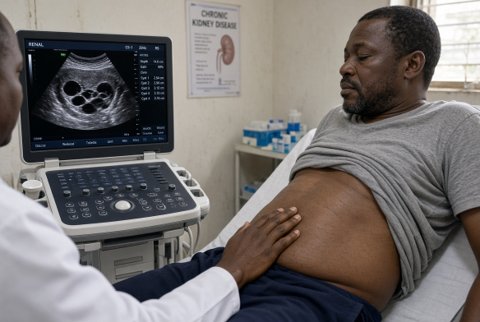
- Ultrasound of the glands and neck as first line imaging for stones, abscess, masses and nodes, with fine needle aspiration cytology or core biopsy of masses (ultrasound guided; sensitivity for malignancy about 80 to 90 percent; core biopsy is more reliable for lymphoma).
- CT with contrast for acute abscess and deep lobe disease; MRI for tumour extent, perineural spread and skull base. MR sialography for strictures. Plain radiograph or cone beam CT for stones (about 80 percent of submandibular stones are radio-opaque).
- Sialendoscopy for diagnosis and treatment of obstructive disease.
- Sjogren syndrome: antinuclear antibody, anti-Ro (SSA) and anti-La (SSB), rheumatoid factor, immunoglobulins, complement, Schirmer test (under 5 mm in 5 minutes), unstimulated salivary flow (under 0.1 mL/min), ocular staining, and labial gland biopsy or parotid ultrasound.
- IgG4 related disease: serum IgG4 (over 135 mg/dL supports), and histology.
- Sarcoidosis: angiotensin converting enzyme, chest radiograph or CT, and biopsy.
- HIV test. Mumps: serum IgM or salivary PCR (notify public health). Interferon gamma release assay and biopsy for tuberculosis.
- Blood glucose, thyroid function, liver function and nutritional screen for sialosis.
- Staging CT or MRI and PET CT for malignancy.
Limitations. Fine needle aspiration can be non-diagnostic and does not always grade tumours; frozen section is unreliable. Biopsy of a parotid mass should not be an open incisional biopsy because of tumour spillage.
Airway compromise or Ludwig angina. Airway control, ceftriaxone 2 g IV every 24 hours plus metronidazole 500 mg IV every 8 hours (or piperacillin with tazobactam 4.5 g IV every 6 to 8 hours), urgent ENT and maxillofacial drainage.
Acute bacterial sialadenitis. Hydration (oral or IV fluids), analgesia, warm compresses, gland massage, sialogogues (sugar free lemon drops or chewing gum), and oral hygiene. Analgesia options are paracetamol 1 g every 4 to 6 hours and ibuprofen 400 mg every 6 to 8 hours with food, avoiding ibuprofen in renal impairment.
Antibiotics: flucloxacillin 500 mg to 1 g orally four times daily for 7 to 10 days (children 12.5 to 25 mg/kg four times daily by age), or co-amoxiclav 625 mg orally three times daily for 7 days. Penicillin allergy: clindamycin 300 to 450 mg orally four times daily or clarithromycin 500 mg twice daily for 7 days.
Severe or systemic illness: flucloxacillin 1 to 2 g IV every 6 hours plus metronidazole 500 mg IV every 8 hours, or co-amoxiclav 1.2 g IV every 8 hours. Add vancomycin when meticillin resistant Staphylococcus aureus is possible (25 to 30 mg/kg IV loading dose, maximum 3 g, then 15 to 20 mg/kg every 8 to 12 hours adjusted to creatinine clearance and levels). Use ultrasound or CT guided aspiration or surgical drainage for abscess.
Salivary stones. Hydration, sialogogues, massage and warm compresses for small stones, with antibiotics when infection coexists. Sialendoscopy with stone removal or lithotripsy for stones under 4 to 7 mm in the parotid or submandibular ducts. Transoral duct incision for distal stones, and gland excision for large intraglandular stones or recurrent infection.
Mumps. Supportive care with paracetamol 1 g orally every 4 to 6 hours, rest, hydration and soft diet; isolation for 5 days after onset of swelling; notify public health. Observe for orchitis (scrotal support, analgesia), meningitis and pancreatitis. Vaccination with two doses of MMR for susceptible contacts.
Juvenile recurrent parotitis. Hydration, massage, antibiotics for acute episodes, and sialendoscopy for frequent episodes; spontaneous resolution after puberty is usual.
Sjogren syndrome.
Management comprises:
- Sugar free sialogogues, saline or saliva substitutes, meticulous oral and dental hygiene with fluoride, and artificial tears.
- Pilocarpine 5 mg orally three to four times daily with meals (maximum 30 mg daily) or cevimeline 30 mg orally three times daily. Avoid pilocarpine in asthma, uncontrolled chronic obstructive pulmonary disease, angle closure glaucoma, and cardiac conduction disease; reduce the dose in hepatic impairment.
- Hydroxychloroquine 200 to 400 mg orally daily (maximum 5 mg/kg actual body weight daily) for systemic features, and rheumatology care.
Annual review for lymphoma risk (persistent gland enlargement, purpura, low C4, cryoglobulins).
IgG4 related disease. Prednisolone 30 to 40 mg orally daily for 2 to 4 weeks then tapered over 3 to 6 months, with rheumatology care and steroid sparing agents (rituximab) for relapse.
Sarcoidosis. Specialist care; prednisolone 20 to 40 mg orally daily for systemic or sight threatening disease.
Sialosis. Treat the cause (diabetes control, nutrition, alcohol cessation, bulimia care, drug change).
Radioiodine sialadenitis. Sialogogues, hydration, anti-inflammatory drugs, and gland massage.
HIV related disease. Antiretroviral therapy; aspiration or sclerotherapy for symptomatic cysts.
Tuberculosis. Isoniazid 5 mg/kg (maximum 300 mg), rifampicin 10 mg/kg (maximum 600 mg), pyrazinamide 25 mg/kg (maximum 2 g) and ethambutol 15 mg/kg daily for 2 months. Then give isoniazid and rifampicin for 4 months, with pyridoxine 10 to 25 mg daily and liver monitoring.
Tumours.
- Benign: superficial or total parotidectomy with facial nerve preservation. Pleomorphic adenoma needs excision with a margin, as rupture risks recurrence; Warthin tumour may be observed in older adults.
- Malignant: surgery with neck dissection by stage and grade, adjuvant radiotherapy for high grade or advanced disease, and chemotherapy or targeted therapy for advanced disease.
- Lymphoma: haematology referral.
Monitoring. Review at 48 to 72 hours for acute infection and at 2 weeks for persistent swelling. Ultrasound surveillance for observed lesions.
Referral and escalation. Immediate for airway compromise, deep neck abscess, or sepsis. ENT or maxillofacial within 2 weeks for any mass, facial nerve palsy, recurrent stones, or persistent swelling beyond 2 weeks after infection. Rheumatology for Sjogren syndrome and IgG4 related disease. Oncology for malignancy.
Admission criteria. Airway threat, abscess needing drainage, sepsis, inability to take oral fluids, or immunosuppression with severe infection.
Discharge and safety netting. Return for fever, spreading redness, facial weakness, difficulty breathing or swallowing, mouth floor swelling, rapid growth of a lump, or pain with numbness.
