








Severe trauma
Also known as: Major trauma, Polytrauma
Severe trauma is injury with a threat to life, limb or function, usually defined by an Injury Severity Score over 15, physiological derangement or a high risk mechanism. Management follows a primary survey of life threats in order of how quickly they kill, with simultaneous treatment, then a secondary survey and definitive care.
Order of the primary survey. Catastrophic external haemorrhage, airway with cervical spine protection, breathing, circulation with haemorrhage control, disability (neurological), and exposure with temperature control. Treat each problem as found before moving on.
Mechanism.
- Blunt: road traffic collisions, falls, assault, crush.
- Penetrating: gunshot, stab, blast.
- Burns, drowning, hypothermia and hyperthermia, electrical injury.
Lethal triad. Hypothermia, acidosis and coagulopathy feed each other and make haemorrhage uncontrollable. Early balanced transfusion, warmth and haemorrhage control break the cycle.
Haemorrhage classes. Class I: under 15% blood volume lost, normal vital signs. Class II: 15 to 30%, tachycardia. Class III: 30 to 40%, hypotension and confusion. Class IV: over 40%, collapse. Shock index over 0.9 predicts need for transfusion.
Triage and trauma team activation.
- Systolic pressure under 90 mmHg.
- Respiratory rate under 10 or over 29 breaths/min.
- Glasgow Coma Scale 13 or less.
- Penetrating injury to the head, neck or torso.
- Flail chest, unstable pelvis, or two or more proximal long bone fractures.
- Amputation proximal to the wrist or ankle, or crush injury.
- Open or depressed skull fracture, or paralysis.
- A fall over 3 metres.
- A major mechanism (ejection, death in the same vehicle, high speed collision, pedestrian or cyclist struck).
Key distinctions. Normal blood pressure does not exclude major bleeding, particularly in young adults, children, athletes and pregnant women. Beta blockers, pacemakers and older age mask tachycardia. A medical event may have caused the injury (hypoglycaemia, seizure, arrhythmia, stroke, intoxication).
Pre-arrival information. Mechanism, time of injury, injuries suspected, vital signs and trend, treatments given (tourniquet time, fluids, drugs), and other casualties.
Primary survey with immediate actions.
- Catastrophic haemorrhage: direct pressure, tourniquet applied 5 to 7.5 cm above a limb wound (note the time; leave in place until surgery, usually safe for up to 2 hours), haemostatic dressing or wound packing.
- Pelvic binder at the level of the greater trochanters for suspected pelvic injury.
- Airway: speak to the patient, look for obstruction, blood, vomit, swelling and burns, and apply manual in-line stabilisation of the cervical spine.
- Breathing: respiratory rate, saturation, chest rise, tracheal position, percussion, air entry, wounds, surgical emphysema.
- Circulation: pulse, blood pressure, capillary refill, skin, neck veins, abdomen, pelvis, femurs and external bleeding.
- Disability: Glasgow Coma Scale, pupils, limb movement, capillary glucose.
- Exposure: remove clothing, log roll with spinal precautions, examine the back and perineum, prevent hypothermia.
Secondary survey. AMPLE history (allergies, medicines, past medical history, last meal, events), head to toe examination including scalp, face, ears, neck, chest, abdomen, pelvis (do not spring the pelvis repeatedly), genitalia, spine, limbs and pulses, rectal and vaginal examination when indicated, and neurological examination.
Red flags.
- Tension pneumothorax (hypotension, absent breath sounds, tracheal deviation, raised jugular venous pressure), massive haemothorax, cardiac tamponade (hypotension, muffled heart sounds, raised venous pressure), open pneumothorax, flail chest.
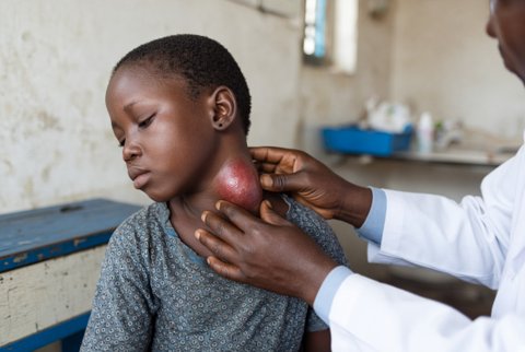
- Expanding neck haematoma, stridor, hard signs of vascular injury (pulsatile bleeding, expanding haematoma, absent pulse, bruit).
- Signs of intra-abdominal bleeding, unstable pelvis, bleeding from the urethra or perineum.
- Glasgow Coma Scale fall, unequal pupils, Cushing response.
- Spinal cord signs and neurogenic shock (hypotension with bradycardia).
- Compartment syndrome (pain out of proportion, tense swelling, pain on passive stretch) and open fracture.
Special groups.
- Children: weight (age based estimate), small airway, intraosseous access early, tachycardia and poor perfusion before hypotension, and consider non-accidental injury.
- Pregnancy: left lateral tilt after 20 weeks, uterine rupture and abruption, foetal monitoring, and anti-D immunoglobulin for rhesus negative mothers.
- Older adults: low energy mechanisms cause serious injury, anticoagulants, comorbidity, and rib fractures.
- Resource limited settings: rely on clinical assessment, basic resuscitation and early transfer, with ultrasound used wherever available.
The differential is of life threatening injuries and of medical causes that mimic or cause trauma.
Thoracic. Tension pneumothorax, open pneumothorax, massive haemothorax, flail chest with pulmonary contusion, cardiac tamponade, blunt cardiac injury, traumatic aortic injury, tracheobronchial injury, oesophageal injury, diaphragmatic rupture, traumatic asphyxia.
Abdominal and pelvic. Liver laceration, splenic rupture, renal injury, mesenteric injury, bowel perforation, pancreatic injury, retroperitoneal haematoma, bladder and urethral injury, unstable pelvic fracture, major vascular injury.
Head and spine. Extradural haematoma, subdural haematoma, traumatic subarachnoid haemorrhage, cerebral contusion, diffuse axonal injury, skull base fracture, cervical spine injury, spinal cord injury with neurogenic shock.
Neck. Laryngeal fracture, carotid or vertebral artery injury, airway transection.
Extremity. Femoral fracture, open fracture, traumatic amputation, arterial injury, compartment syndrome, crush syndrome with rhabdomyolysis, fat embolism.
Environmental. Burns, inhalation injury, drowning, hypothermia, heat stroke, electrical injury, blast injury.
Medical causes of injury or collapse. Hypoglycaemia, seizure, stroke, subarachnoid haemorrhage, myocardial infarction, arrhythmia, syncope, drug or alcohol intoxication, and pregnancy related collapse.
Non-accidental. Child abuse, intimate partner violence, elder abuse.
Resuscitation phase.
- Extended focused assessment with sonography (heart, hepatorenal and splenorenal spaces, pelvis, lungs for pneumothorax and haemothorax): quick, repeatable, and shows free fluid or pericardial blood. A negative result does not exclude injury, particularly retroperitoneal and hollow viscus injury.
- Chest and pelvis radiographs if they do not delay resuscitation.
- Blood: gas with lactate and base deficit (base deficit worse than minus 6 mmol/L signals severe shock), haemoglobin (may be normal early), coagulation with fibrinogen, viscoelastic testing where available, group and crossmatch.
- Also electrolytes, ionised calcium, creatinine, creatine kinase, glucose, blood alcohol, pregnancy test, troponin and ECG with chest trauma.
Definitive imaging (once stable or responding).
- Whole body contrast CT (head to pelvis, with CT angiography of the chest, abdomen and pelvis) for patients who are stable or respond to resuscitation. Unstable patients with a positive ultrasound go to theatre or interventional radiology.
- CT head for Glasgow Coma Scale under 13 at any time, under 15 at 2 hours, suspected open or depressed skull fracture, signs of basal skull fracture, or more than one vomit.
- Also CT head for age 65 or over with loss of consciousness or amnesia, dangerous mechanism, or anticoagulation (within 8 hours, and within 1 hour when other high risk features are present).
- CT cervical spine for Glasgow Coma Scale under 15, age 65 or over, dangerous mechanism, focal neurological deficit, paraesthesia, or inability to assess.
- Clearance of the cervical spine without imaging only when the patient is alert, sober, has no distracting injury, no midline tenderness and full painless neck rotation.
- Angiography for neck and extremity vascular injury. Cystourethrography for suspected urethral injury (before urethral catheterisation with blood at the meatus). Echocardiography for suspected blunt cardiac or valvular injury.
Tertiary survey. Repeat full examination and review of imaging within 24 hours, since 2 to 10% of injuries are missed initially.
Limitations. Initial haemoglobin and blood pressure underestimate bleeding. Ultrasound is operator dependent. CT carries radiation exposure, which is justified in severe trauma but limited in children and pregnancy to targeted studies.
Airway and cervical spine. Jaw thrust, suction, airway adjuncts. Intubate for airway obstruction, Glasgow Coma Scale 8 or less, inadequate ventilation, severe facial or neck injury, or impending airway loss, using manual in-line stabilisation, preoxygenation and a bougie.
Rapid sequence induction: ketamine 1 to 2 mg/kg IV (0.5 to 1 mg/kg in shock) or etomidate 0.3 mg/kg IV, with rocuronium 1.2 mg/kg IV or suxamethonium 1.5 mg/kg IV.
Avoid suxamethonium with hyperkalaemia, crush injury beyond 24 hours, burns beyond 24 hours, denervation and malignant hyperthermia risk. Monitor blood pressure, which may fall at induction, so resuscitate before induction where possible. Surgical airway if intubation fails.
Breathing.
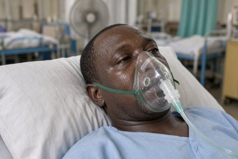
- Oxygen 15 L/min by reservoir mask, then titrate to saturation 94 to 98%.
- Tension pneumothorax: immediate finger or open thoracostomy in the fifth intercostal space anterior to the mid axillary line, then a chest drain (28 Fr or larger for haemothorax, 20 to 24 Fr for pneumothorax). Needle decompression is a temporary step with a high failure rate.
- Open pneumothorax: three sided occlusive dressing, then a chest drain at a separate site.
- Massive haemothorax: drain and transfuse. Immediate drainage over 1500 mL or ongoing drainage over 200 mL/hour for 2 to 4 hours is an indication for thoracotomy.
- Flail chest: analgesia (regional block), oxygen, and ventilation if respiratory failure.
Circulation: damage control resuscitation.
- Two large bore cannulae or intraosseous access, and activation of massive transfusion when ongoing bleeding is expected.
- Give red cells, plasma and platelets in near equal proportions (about 1:1:1), warmed, and limit crystalloid to under 1 L.
- Aim for a systolic pressure of 80 to 90 mmHg until bleeding is controlled (permissive hypotension), and avoid it with head injury or spinal injury, where a systolic pressure over 110 mmHg is wanted.
- Tranexamic acid 1 g IV over 10 minutes within 3 hours of injury, then 1 g IV over 8 hours; monitor for thrombosis and seizures; reduce in kidney impairment. Children: 15 mg/kg IV (maximum 1 g), then 2 mg/kg/hour.
- Calcium chloride 10% 10 mL IV after every 4 units of red cells, keep ionised calcium over 1.1 mmol/L. Fibrinogen concentrate 2 to 4 g IV or cryoprecipitate when under 1.5 g/L. Platelets to over 50 x 10^9/L (over 100 x 10^9/L in head injury). Warm the patient to over 36 °C.
- Reverse anticoagulants:
- warfarin with 4 factor prothrombin complex concentrate 25 to 50 units/kg IV and vitamin K 5 to 10 mg IV
- dabigatran with idarucizumab 5 g IV
- factor Xa inhibitors with prothrombin complex concentrate 50 units/kg IV or the specific antidote from the product label
- Haemorrhage control: tourniquet, packing, pelvic binder, resuscitative thoracotomy (penetrating chest trauma with arrest in the last minutes), laparotomy, preperitoneal packing, interventional radiology embolisation. Specialist teams may use endovascular balloon occlusion of the aorta.
- Tamponade: pericardiocentesis or thoracotomy.
- Neurogenic shock: restore volume, then noradrenaline 0.05 to 0.5 micrograms/kg/min IV titrated to a mean pressure of 85 to 90 mmHg for the first days after cord injury (evidence is limited), and atropine 500 micrograms IV for bradycardia.
Disability.
- Head injury: treat hypoxia and hypotension (the main secondary injuries), head up 30 degrees, PaCO2 4.5 to 5.0 kPa, glucose, and temperature control.
- Raised pressure signs: mannitol 0.5 to 1 g/kg IV over 15 to 20 minutes or 3% sodium chloride 150 to 250 mL IV over 10 to 20 minutes, with urgent neurosurgical review.
- Monitor sodium every 4 to 6 hours (stop if over 160 mmol/L), and osmolality (stop mannitol if over 320 mOsm/kg).
- Seizure: lorazepam 4 mg IV, repeat once after 10 minutes.
- Spinal injury: log roll and immobilise, maintain perfusion, and arrange urgent spinal assessment. High dose corticosteroids are not advised.
Analgesia.
- Fentanyl 25 to 50 micrograms IV every 3 to 5 minutes titrated to effect (reduce in shock, older age and head injury; monitor respiration and sedation; maximum about 200 micrograms before reassessment).
- Alternatively, ketamine 0.1 to 0.3 mg/kg IV as an analgesic dose.
- Paracetamol 1 g IV every 6 hours (maximum 4 g daily; 3 g daily if weight under 50 kg, malnutrition or liver disease).
- Regional anaesthesia for rib and femoral fractures.
Open fractures and wounds.
- Amoxicillin with clavulanic acid 1.2 g IV every 8 hours, given within 1 hour of injury, continued for 24 hours after wound closure (extended to 72 hours or until soft tissue cover for severe contaminated injuries).
- Alternative: cefazolin 2 g IV every 8 hours.
- Add gentamicin 5 mg/kg IV once for severe contaminated injuries under specialist direction, with monitoring of creatinine and levels.
- Tetanus: tetanus toxoid 0.5 mL IM when the last dose was over 10 years ago (over 5 years for dirty wounds) or immunisation is unknown or incomplete.
- Add tetanus immunoglobulin 250 units IM (500 units when over 24 hours since injury or with heavy contamination) for high risk wounds with unknown or incomplete immunisation.
- Irrigate and debride early, splint, and cover with a sterile dressing. Realign and splint fractures, and check distal pulses before and after.
Compartment syndrome. Remove constricting dressings, keep the limb at heart level, give analgesia, and arrange emergency fasciotomy for clinical diagnosis or a pressure within 30 mmHg of the diastolic pressure.
Crush injury. Isotonic crystalloid 1 to 1.5 L/hour before release if possible, then 500 mL/hour, aiming for urine output over 200 mL/hour; monitor potassium, creatine kinase and ECG; treat hyperkalaemia with calcium chloride 10% 10 mL IV and insulin 10 units with glucose 25 g.
Special measures.
- Prevent hypothermia (warm fluids, blankets, warm room).
- Gastric and bladder decompression when indicated.
- Venous thromboembolism prophylaxis with enoxaparin 40 mg subcutaneously once daily, started when bleeding is controlled, usually within 24 to 72 hours.
- Reduce enoxaparin to 20 mg daily if CrCl 15 to 30 mL/min. Use unfractionated heparin 5000 units subcutaneously every 8 to 12 hours if CrCl under 15 mL/min.
- Use mechanical prophylaxis until then.
Monitoring. Continuous ECG, saturation, capnography, arterial pressure, core temperature, hourly urine output, lactate every 1 to 2 hours, repeat gas, haemoglobin, coagulation and calcium, neurological observations, serial abdominal and limb examination, and repeat imaging for changes.
Referral and escalation. Trauma team leader, general or trauma surgery, orthopaedics, neurosurgery, anaesthesia and critical care, interventional radiology, vascular surgery, plastic surgery, ENT or maxillofacial, and obstetrics. Transfer unstable patients and those needing specialist care to a major trauma centre, with a clear handover.
Admission criteria. All patients with severe trauma are admitted to critical care, theatre or a high dependency unit. Patients with minor injury, a normal examination and normal imaging may go home after a tertiary survey, observation and a responsible adult.
Discharge and safety netting. Discharge only after a tertiary survey and review of all imaging. Patients are told to return for worsening headache, vomiting, drowsiness, confusion, weakness, new abdominal pain, shoulder tip pain, breathlessness, chest pain, black stools, haematuria, limb swelling or numbness, or fever. Arrange fracture clinic, physiotherapy, psychological support (post-traumatic stress), safeguarding where non-accidental injury is suspected, and advice on driving and return to work.
