








Image-Guided Biopsy
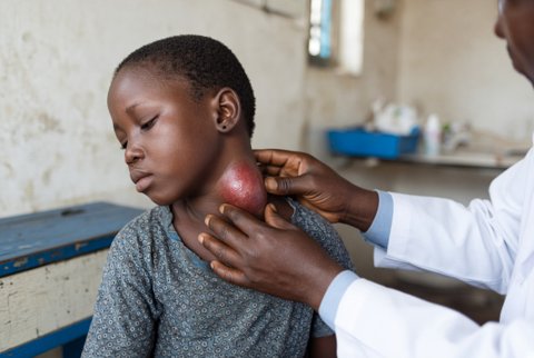
Biopsy under image control is the sampling of tissue from a lesion, organ or lymph node using a needle placed under ultrasound, CT, fluoroscopy or MR control.
Fine needle aspiration (21 to 25 gauge) yields cells for cytology, and core biopsy (14 to 20 gauge, automated spring loaded or semi-automatic needles, coaxial systems) yields tissue cores for histology, immunohistochemistry and molecular tests.
Ultrasound is preferred for superficial organs, liver, kidney, thyroid, breast, lymph nodes, pleura and pelvis because it shows the needle in real time without radiation. CT is used for lung, mediastinum, bone, retroperitoneum, deep abdominal and pelvic lesions, using intermittent scanning or fluoroscopy mode.
Fluoroscopy is used for bone, spine and some chest lesions, and MR for lesions seen only on MR (prostate, breast, brain, musculoskeletal). Biopsy under endoscopic ultrasound reaches pancreas, nodes and gastric wall lesions. The planned path avoids vessels, bowel, lung, nerves and ducts.
Biopsy under image control replaced much surgical biopsy because it is faster, cheaper, and has fewer complications. Diagnostic yield is 85 to 98% for core biopsy of most solid organs (liver about 95%, lung 85 to 95%, bone 70 to 90%, kidney over 90%, lymph node over 90% with core biopsy), and major complications occur in under 1 to 3%.
Tissue diagnosis changes treatment: it defines malignancy, type, grade and receptor or mutation status, confirms infection and directs targeted therapy.
