











Neuroanatomy Imaging and Surface Orientation
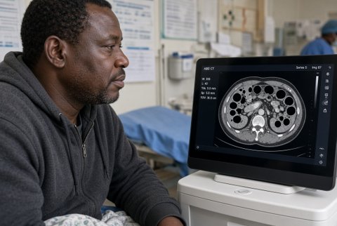
This final section translates the structural anatomy into the imaging planes used in everyday clinical practice, since recognising a structure on a scan is the practical skill that validates and reinforces textbook neuroanatomy knowledge.
- The standard, most commonly acquired imaging plane for both CT and MRI, viewed by convention as though looking up from the patient's feet (radiological convention: the patient's right side appears on the image's left side), a series of axial slices, read from inferior to superior, is the default way most students and clinicians first learn to recognise brain structures, since a single well-chosen axial slice can display several structures simultaneously (e.g., a slice through the basal ganglia typically also captures the thalamus, internal capsule, and lateral ventricles together).
- Slices oriented perpendicular to the axial plane, viewed as though facing the patient directly (again by radiological convention, the patient's right appears on the image's left), particularly useful for assessing structures with a predominantly vertical orientation or symmetry (e.g., comparing the two hippocampi, or assessing the corpus callosum and lateral ventricles' relationship to the midline), and for structures like the pituitary gland/sella turcica region, where the coronal plane best demonstrates the relationship to the optic chiasm above and the cavernous sinuses laterally.
- Slices oriented in the anteroposterior plane, dividing the brain into left/right portions, the midsagittal slice is particularly valuable, displaying the corpus callosum, the third ventricle, the brainstem's full length in profile, the cerebellum, and the pituitary/sella turcica region all within a single image, a classic "orientation" slice frequently used to introduce the brain's overall midline architecture before moving to axial or coronal detail.
- CT (computed tomography) remains the first-line imaging modality in acute settings (particularly trauma and suspected haemorrhage), given its speed, wide availability, and excellent sensitivity for acute blood (which appears hyperdense/bright relative to normal brain tissue), the imaging correlate of the haemorrhage patterns and 16: epidural haemorrhage appears as a lentiform (biconvex), suture-limited hyperdensity; subdural haemorrhage appears as a crescent-shaped hyperdensity, not suture-limited, often layering along the brain's contour; subarachnoid haemorrhage appears as hyperdensity tracking within the sulci/cisterns/ventricular system rather than forming a discrete collection.
- CT also readily demonstrates the ventricular system's size/shape (for hydrocephalus assessment), skull fractures, and, with lower sensitivity than MRI but adequate for urgent triage, established (though not hyperacute) ischaemic stroke territory (as a hypodense region corresponding to the affected vascular territory).
- MRI (magnetic resonance imaging) offers superior soft tissue contrast and multiplanar capability compared to CT, making it the preferred modality for detailed structural assessment, early ischaemic stroke detection (via diffusion-weighted sequences, sensitive to the cytotoxic oedema of acute ischaemia within minutes to hours, well before CT would show any change), and posterior fossa/brainstem imaging (where CT is limited by bone-related artefact, given the dense surrounding skull base), the fundamental structural anatomy (cortical gyri/sulci, white matter tracts, ventricles, brainstem, cerebellum, spinal cord) is most clearly and fully visualised on MRI, making familiarity with normal MRI anatomy the single most valuable applied skill for consolidating this content's structural material.
- The lateral ventricles are readily identified on axial imaging as paired, curved, fluid-filled (CT: hypodense; standard MRI T2: hyperdense) structures, with their frontal horns anteriorly and (at a slightly higher or lower slice, depending on the precise level) their atria/occipital horns posteriorly and temporal horns inferolaterally, their size and symmetry are a routine, essential part of every brain scan assessment, given their sensitivity to both hydrocephalus (enlargement) and mass effect/midline shift (asymmetric displacement).
- The third ventricle appears as a thin, midline, slit-like structure at an axial level through the thalami (visible between the two thalami), or clearly on midsagittal imaging.
- The fourth ventricle appears as a small, midline, diamond-shaped structure at the level of the pons/cerebellum, best appreciated on axial imaging through the posterior fossa or on midsagittal imaging.
- The cerebral aqueduct, given its small calibre, is not always clearly resolved on standard imaging but its patency (or obstruction) can be inferred from the pattern of ventricular dilatation above vs below it.
- Best appreciated on an axial slice through the level of the thalami/third ventricle, the caudate nucleus head is seen bulging into the lateral wall of the frontal horn; the putamen lies lateral to the globus pallidus, both together forming the lentiform nucleus, itself lateral to the internal capsule's posterior limb; the internal capsule (both limbs and the genu) is typically well seen at this same level, sandwiched between the caudate/thalamus medially and the lentiform nucleus laterally, a single well-chosen axial slice at this level is one of the highest-yield images in the entire brain for consolidating the spatial relationships.
- Appears as paired, ovoid grey matter masses flanking the third ventricle, at the same general axial level as the basal ganglia, individual thalamic nuclei are not routinely distinguishable on standard clinical imaging (given their small size and subtle contrast differences), but the thalamus's overall size, symmetry, and relationship to the adjacent internal capsule/ventricles remain clinically important, particularly for localising small deep (lacunar) infarcts or haemorrhages in this region.
- On axial imaging through the posterior fossa, the brainstem's characteristic shape changes distinctly at each level, providing a reliable way to identify which level (midbrain, pons, or medulla) a given slice represents: the midbrain shows a characteristic "Mickey Mouse" or owl-like appearance (the cerebral peduncles forming the "ears," the tectum/colliculi posteriorly, the cerebral aqueduct centrally); the pons appears as a larger, more rounded/oval structure with the fourth ventricle posteriorly and the middle cerebellar peduncles extending laterally toward the cerebellum; the medulla appears smaller and more elongated, with the pyramids visible anteriorly and (at its lower extent) beginning to resemble the spinal cord's more circular cross-sectional shape, this level-specific shape recognition is the essential imaging skill underlying the "crossed syndrome" localisation principle established in Sections 7, 12, and 16, since correctly identifying the axial level shown is the first step in correlating an imaging abnormality with a specific combination of expected cranial nerve nuclei.
- Occupies the posterior fossa, readily identified on axial imaging by its characteristic folded, layered (folia) surface texture, distinct from the smoother appearance of the overlying cerebral hemispheres, with the vermis forming the midline connection between the two hemispheres: the vermis vs hemisphere distinction relevant to lesion localisation (Section 8, 16) is directly visualisable, particularly on coronal or axial imaging showing both structures simultaneously.
- Recognising the characteristic shape and location of an infarct on imaging allows direct inference of the occluded vessel, applying the territorial anatomy: an MCA territory infarct produces a wedge-shaped area of abnormality involving the lateral hemisphere surface (cortex and adjacent white matter), often with a sharp, sparing border along the medial hemisphere surface (the ACA/MCA watershed boundary); an ACA territory infarct produces a corresponding wedge-shaped abnormality on the medial hemisphere surface; a PCA territory infarct produces abnormality involving the occipital lobe and/or medial temporal lobe; a small, discrete, rounded lesion within the internal capsule/basal ganglia/thalamus (rather than a larger wedge-shaped cortical territory) suggests a lacunar infarct, reflecting occlusion of a single small perforating vessel, this pattern-recognition skill, connecting infarct shape/location on imaging to the underlying vascular anatomy, is a direct, practical extension of the territorial knowledge.
- Best assessed on sagittal MRI for an overview of the cord's full length, alignment, and any focal signal abnormality or compression (e.g., from disc herniation, tumour, or vertebral pathology) relative to specific vertebral levels, axial imaging through the cord at a specific level demonstrates its cross-sectional grey/white matter organisation, useful for localising the precise location of an intramedullary lesion (e.g., central vs eccentric, anterior vs posterior) and directly correlating with the classic lesion patterns (a central lesion suggesting syringomyelia/central cord syndrome; an anterior lesion suggesting anterior spinal artery territory involvement; a unilateral lesion suggesting a process consistent with Brown-Séquard-type hemisection).
