









Leishmania
Leishmania comprises a genus of obligate intracellular protozoan parasites transmitted by sandflies, producing three principal clinical syndromes depending on the infecting species, the parasite's tissue tropism, and the host immune response.
Visceral leishmaniasis (kala-azar) is the most severe form. It is caused principally by Leishmania donovani (Indian subcontinent, East Africa) and Leishmania infantum (Mediterranean basin, Latin America, where it is also referred to as Leishmania chagasi).
The parasite disseminates to and replicates within the reticuloendothelial system, infecting macrophages throughout the spleen, liver, bone marrow, and lymph nodes. Untreated visceral leishmaniasis is fatal in the large majority of symptomatic cases, making it the deadliest of the three forms.
Classic features of visceral leishmaniasis are:
- Prolonged fever.
- Marked splenomegaly (often massive) with lesser hepatomegaly.
- Progressive pancytopenia, from marrow infiltration, splenic sequestration, and hypersplenism.
- Weight loss and wasting.
- Hypergammaglobulinaemia with a reversed albumin-to-globulin ratio.
Skin darkening is a recognised feature in some endemic populations, particularly on the Indian subcontinent. It gives kala-azar its name, meaning "black fever" or "black sickness".
Cutaneous leishmaniasis is the most common clinical form worldwide. Many species cause it, depending on region:
- Leishmania major and Leishmania tropica (Old World, Middle East, North Africa, Central Asia).
- Leishmania mexicana and Leishmania braziliensis (New World, Central and South America). Braziliensis is more closely associated with mucocutaneous disease.
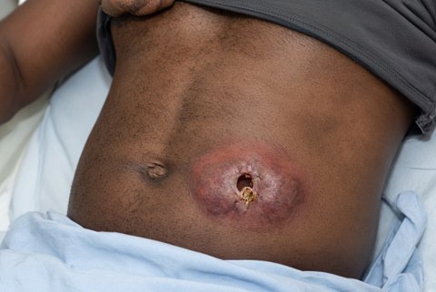
Infection remains localised to the skin at or near the sandfly bite site. A papule enlarges and typically ulcerates over weeks to months. The ulcer is painless in most cases, with raised, indurated borders often described as volcano-shaped. It heals slowly over months to years, leaving a disfiguring scar.
Multiple bites can cause multiple lesions. Satellite lesions or regional lymphadenopathy may develop.
Mucocutaneous leishmaniasis is a New World syndrome, most strongly associated with Leishmania braziliensis (and related species within the Viannia subgenus). Parasites spread from the primary cutaneous lesion by lymphatic or haematogenous dissemination to the nasopharyngeal and oral mucosa. This typically presents months to years after apparent healing of the original skin lesion.
Progressive, destructive granulomatous inflammation of the nasal septum, palate, and pharyngeal tissue produces severe tissue destruction. This includes nasal septal perforation and collapse, producing the classically described "tapir nose" or "espundia" deformity.
The disease can extend to the larynx, with risk of airway compromise and disfigurement. Unlike cutaneous disease, mucocutaneous leishmaniasis does not spontaneously resolve and requires treatment.
