










Free Living Amoebae
Naegleria fowleri is a thermophilic free-living amoeba found in warm freshwater environments. These include lakes, hot springs, poorly chlorinated swimming pools, and warm tap water in some regions. It causes primary amoebic meningoencephalitis, an extremely rare but almost uniformly fatal acute CNS infection.
Unlike the exclusively human-parasitic organisms, Naegleria fowleri exists and thrives independently in the environment. Human infection is an incidental, dead-end event that does not contribute to the organism's normal life cycle.
It exists in three forms:
- a hardy environmental cyst
- a feeding and dividing trophozoite (amoeboid form)
- a transient, non-feeding, flagellate form that the trophozoite can convert into under adverse environmental conditions such as low nutrient availability
The flagellate form is a distinguishing biological feature not shared by Acanthamoeba or Balamuthia.
Acanthamoeba species are also free-living amoebae. They are ubiquitous in soil, dust, freshwater, and even tap water and improperly maintained contact lens solutions.
They exist in only two forms, a trophozoite and a highly resistant double-walled cyst. The cyst is more resistant than the Naegleria cyst, contributing to its notorious resistance to standard disinfection.
Acanthamoeba causes two principal human disease syndromes. The populations at risk and the pathophysiology differ markedly.
Balamuthia mandrillaris is a free-living amoeba found in soil. It was identified relatively recently compared with Naegleria and Acanthamoeba. Like Acanthamoeba, it exists as a trophozoite and a resistant cyst without a flagellate stage.
Balamuthia causes a chronic granulomatous encephalitis with substantial clinical overlap with Acanthamoeba CNS disease. It also affects immunocompetent individuals more often than Acanthamoeba GAE does.
It characteristically produces skin lesions that can precede CNS involvement by weeks to months. This offers a potential window for earlier recognition if suspected.
All three organisms share a fundamentally environmental, saprophytic existence, feeding on bacteria and organic debris in soil and water. They do not require a host to complete their life cycle, which distinguishes them from every other protozoan covered in this module.
Naegleria fowleri is specifically associated with warm freshwater and is not found in seawater. Optimal growth occurs at higher temperatures, up to around 45 degrees Celsius. This explains its association with hot springs, warm lakes in summer months, and inadequately chlorinated or heated recreational water.
Acanthamoeba and Balamuthia are more widely distributed in soil and are also found in freshwater. Acanthamoeba is also found in a broad range of man-made environments, including tap water, air conditioning systems, and contact lens equipment. This is directly relevant to its most common clinical association.
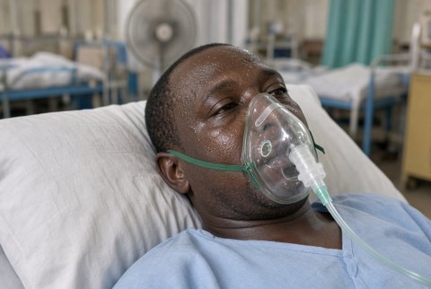
Naegleria fowleri causes primary amoebic meningoencephalitis (PAM), an acute, fulminant infection that progresses rapidly over days. It typically presents in previously healthy children and young adults with a recent history of freshwater exposure (swimming, diving, or nasal irrigation with contaminated water).
Features are severe headache, fever, nausea, and vomiting, with rapid progression to meningismus, altered mental status, seizures, and coma. The clinical tempo and severity closely mimic acute bacterial meningitis.
Mortality exceeds 95 percent even with treatment. This reflects both the rapidity of disease progression and the limited efficacy of available therapy once established.
Acanthamoeba causes granulomatous amoebic encephalitis (GAE), a subacute to chronic CNS infection evolving over weeks to months. It occurs predominantly in immunocompromised individuals (including those with HIV/AIDS, on immunosuppressive therapy, or with chronic debilitating illness), in contrast to the healthy host typical of Naegleria PAM.
Presentation includes headache, focal neurological deficits, seizures, and altered mental status. Imaging typically shows single or multiple space-occupying lesions. The chronic course frequently leads to diagnostic delay or misdiagnosis as tumour, abscess, or other granulomatous disease before amoebic infection is considered.
Balamuthia mandrillaris also causes a granulomatous encephalitis clinically similar to Acanthamoeba GAE, subacute to chronic in course. It affects a broader population, including immunocompetent individuals. Children and older adults appear disproportionately represented among reported cases relative to Acanthamoeba GAE.
As with Acanthamoeba GAE, prognosis is poor and mortality is similarly very high. Case reports of survival with early diagnosis and aggressive combination therapy exist for both organisms.
Acanthamoeba keratitis is a distinct and important disease entity, a sight-threatening infection of the cornea strongly associated with contact lens use. Risk factors:
- exposure to contaminated water while wearing lenses (swimming, showering, or rinsing lenses in tap water or homemade saline rather than sterile solution)
- poor lens hygiene
- use of inadequately disinfecting contact lens solutions
It presents with severe ocular pain, photophobia, redness, and blurred vision. The pain is often disproportionate to visible clinical signs, a classically noted feature. A ring-shaped corneal infiltrate appears in more advanced disease.
It is frequently misdiagnosed initially as herpes simplex keratitis or bacterial/fungal keratitis, given overlapping features. This delay worsens outcomes. Acanthamoeba keratitis requires prolonged, specific antiamoebic topical therapy. Otherwise it can progress to corneal scarring, perforation, and significant visual loss.
Naegleria fowleri and Balamuthia mandrillaris are not associated with keratitis. This is a disease specific to Acanthamoeba among the free-living amoebae.
Naegleria fowleri enters the human host through the nasal mucosa. This typically happens during activities that force water up the nose (diving, jumping into water, water sports, or nasal irrigation with inadequately treated water).
Trophozoites then migrate along the olfactory nerve fibres, crossing the cribriform plate to reach the olfactory bulbs and then the brain directly. This short and efficient anatomical route contributes to the rapid, fulminant course of PAM. Infection is not acquired by drinking contaminated water; nasal instillation is specifically required.
Acanthamoeba causing GAE is thought to enter primarily via the respiratory tract or through breaks in the skin, with haematogenous dissemination to the CNS. This route is consistent with its more common occurrence in the setting of systemic immunosuppression.
Acanthamoeba keratitis, by contrast, results from direct corneal inoculation, typically via contact lenses or corneal trauma. It does not involve CNS spread as an intermediate step.
Balamuthia mandrillaris is thought to enter through breaks in the skin (consistent with the skin lesions that often precede or accompany CNS disease) or via the respiratory tract. Haematogenous spread to the central nervous system follows, broadly analogous to the presumed route for Acanthamoeba GAE.
Diagnosis of primary amoebic meningoencephalitis relies on prompt recognition, given the extremely rapid clinical course. Cerebrospinal fluid typically shows a neutrophil-predominant pleocytosis, elevated protein, and low glucose. This closely mimics bacterial meningitis and can misdirect initial management if amoebic infection is not specifically considered.
Definitive diagnosis requires direct visualisation of motile trophozoites on wet mount microscopy of fresh, unrefrigerated cerebrospinal fluid. This is a critical technical point, since refrigeration or delay markedly reduces the ability to observe characteristic amoeboid motility. PCR supports the diagnosis where available. It offers more rapid and specific confirmation and is increasingly the preferred approach.
Diagnosis of granulomatous amoebic encephalitis (Acanthamoeba or Balamuthia) is often made on brain biopsy. The chronic, indolent presentation frequently mimics other space-occupying or granulomatous CNS processes. Histopathology shows trophozoites and cysts within areas of granulomatous inflammation and necrosis.
PCR and, for Balamuthia, serology are increasingly used adjuncts. Diagnosis is frequently made late or only at autopsy, given the rarity and nonspecific presentation of these infections.
Diagnosis of Acanthamoeba keratitis relies on corneal scraping for microscopy (identifying trophozoites or the characteristic double-walled cysts). Culture is on non-nutrient agar seeded with bacteria, which the amoeba feeds upon.
Confocal microscopy of the living cornea and PCR are increasingly used as adjunctive or alternative diagnostic tools. They are particularly valuable given the clinical overlap with other infectious keratitis syndromes and the importance of prompt, correct diagnosis in preventing vision-threatening progression.
