











Post-Void Residual Measurement
The post-void residual is the volume of urine left in the bladder after the patient has passed urine as completely as they can. It shows how well the bladder empties, and it identifies retention before it causes pain, infection, stones or kidney damage.
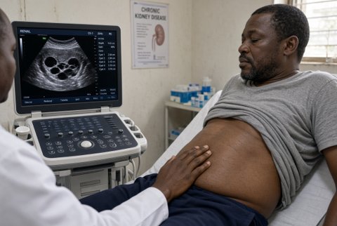
It is measured by ultrasound (a bladder scanner or a conventional ultrasound machine) or by catheterisation. The scan is done within 5 to 10 minutes of voiding, and takes about a minute. A portable bladder scanner uses automated three dimensional or multiplane measurements to give a volume.
With a conventional probe, volume is calculated from three bladder diameters using a simple formula (height x width x depth x about 0.52). Catheterisation gives an exact volume but is invasive, and it carries risks of infection, pain and urethral injury.
The accuracy of a bladder scan is within about 15 to 20% of the true volume, with the greatest error at small volumes, in women with pelvic masses or ascites, and in obese patients. Residual volume varies from one void to the next, and one measurement may mislead.
Chronic retention affects up to 10% of older men and a similar proportion of women with neurological disease or after pelvic surgery. In older men with lower urinary tract symptoms, about 10 to 15% have a residual of over 100 mL.
Indications.
- Lower urinary tract symptoms: hesitancy, weak stream, incomplete emptying, overflow incontinence, and recurrent urinary infection.
- Neurogenic bladder (spinal cord injury, multiple sclerosis, diabetes, spina bifida), including surveillance.
- After surgery, anaesthesia, or childbirth, with suspected retention.
- Medication that can impair emptying (anticholinergic, opioid, and some antidepressant and antipsychotic drugs).
- Hydronephrosis, raised creatinine, or recurrent infection of unknown cause.
- After surgery on the prostate, urethra or pelvic floor, and after incontinence procedures.
- Before starting treatment for overactive bladder, to exclude retention.
- Suspected urethral stricture and bladder outlet obstruction.
Not indicated for diagnosing the cause of the retention, which needs further tests.
Contraindications and precautions.
- No contraindication for ultrasound. Open wounds, dressings, or a catheter in place limit scanning.
- Catheterisation is avoided where there is urethral injury (blood at the meatus after trauma), and is done with care in a patient with a stricture or recent urethral surgery.
- Scans in women with a pelvic mass, large fibroids, ovarian cysts or ascites can give false high readings.
- Patients with a very full bladder or in distress are not made to wait for a scan.
Preparation and technique.
The patient is asked to void as completely as possible, in a comfortable position and privacy, and scanning is done straight after (within 10 minutes). The probe is placed on the suprapubic area with gel and aimed at the bladder, and the scanner outlines the bladder in several planes.
The scan is repeated if the reading seems doubtful, and the bladder shape and the position of the cursor are checked on the screen. When two or three measurements differ, the lowest, or the average, is used depending on the local routine, and a repeat on another day is done for borderline results.
