










Renal Biopsy
Renal biopsy removes a small sample of kidney tissue, usually with a needle, for examination by light microscopy, immunofluorescence and electron microscopy. It gives the diagnosis, the degree of activity and scarring, and the outlook for many kidney diseases that cannot be named by blood and urine tests alone.
A percutaneous biopsy of a native kidney is done with the patient lying prone, using real time ultrasound to find the lower pole of the kidney, where the cortex is thickest and large vessels are fewest. A spring loaded 16 to 18 gauge needle takes 2 to 3 cores. Biopsy of a transplant kidney is done in the iliac fossa. When the percutaneous approach is risky (single kidney, bleeding disorder, obesity, uncontrolled hypertension), a transjugular approach or open or laparoscopic biopsy can be used.
The procedure takes 15 to 30 minutes, and observation lasts 6 to 24 hours. An adequate sample has at least 10 to 15 glomeruli for light microscopy, and gives a diagnosis in over 95% of cases. A biopsy changes the diagnosis or management in about 40 to 50% of patients with nephrotic syndrome or unexplained renal impairment.
Indications.
- Nephrotic syndrome in adults, and in children when steroid resistant or atypical.
- Nephritic syndrome, rapidly progressive glomerulonephritis, or unexplained acute kidney injury that does not improve.
- Persistent non-visible haematuria with proteinuria, or isolated proteinuria over about 1 g per day, with glomerular disease suspected.
- Chronic kidney disease of unknown cause with preserved kidney size.
- Suspected lupus nephritis, vasculitis, or kidney involvement in systemic disease (myeloma, amyloidosis, diabetes with atypical features).
- Kidney transplant dysfunction, to separate rejection, recurrent disease, drug toxicity and infection.
- Suspected acute interstitial nephritis, and diagnosis in a familial kidney disease (such as Alport syndrome).
- Assessment of disease activity before changing treatment.
Not indicated when the diagnosis is already clear (for example, diabetic nephropathy in a typical patient) and the result will not change management.
Contraindications and precautions.
- Absolute: uncorrected bleeding disorder, an uncooperative patient who cannot hold the breath or lie still, and a solitary native kidney (unless there is no other route and the benefit is clear).
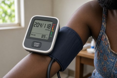
- Relative: uncontrolled hypertension (target under about 140/90 mmHg), small, shrunken, scarred kidneys (under 9 cm, which are risky and give little information), active urinary or skin infection over the site, hydronephrosis, kidney cysts or tumour, severe anaemia, and obesity.
- Anticoagulant and antiplatelet drugs are stopped for the right time before biopsy, and bleeding tests (platelet count, prothrombin time, activated partial thromboplastin time) are checked, with a platelet count of at least 100 x 10⁹/L preferred.
- Uraemic patients have platelet dysfunction, which can be improved by desmopressin or dialysis before the biopsy.
- A blood group and a sample for cross-match are available.
Preparation and technique.
Consent covers the risks of bleeding, pain, the need for transfusion, and rarely, surgery or loss of the kidney. Blood pressure is controlled, and an intravenous line is placed. Ultrasound confirms two kidneys, size and position, and the site is marked.
The skin is cleaned, local anaesthetic is infiltrated down to the kidney capsule, and the patient holds the breath during needle passage, which is done at the end of inspiration or expiration as practised.
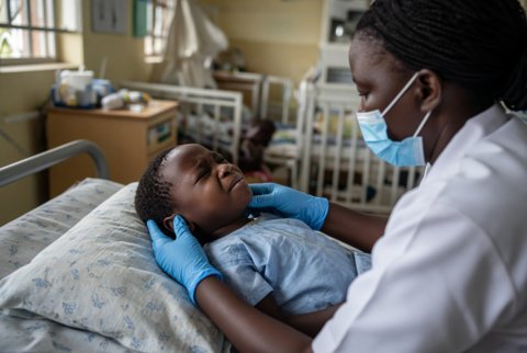
The core is checked under a dissecting microscope or loupe for glomeruli, and divided for light microscopy (formalin), immunofluorescence (frozen or special medium) and electron microscopy (glutaraldehyde). After the biopsy, the patient lies supine at rest for 4 to 6 hours or more, with blood pressure, pulse and urine colour monitored.
