










Localised itching
Also known as: Localized itching
Localised itching is pruritus confined to one body region. Itch on normal skin in a nerve distribution suggests neuropathic itch, while itch with visible lesions usually reflects local inflammation, infection or infestation. It covers the trunk, limbs, face, hands and feet.
Classification by skin findings. Itch on visibly abnormal skin, itch on normal skin, and itch with chronic scratch lesions (lichen simplex chronicus, prurigo nodules).
Classification by mechanism. Inflammatory, infective or infestation, irritant or allergic contact, neuropathic, systemic disease with a local manifestation, and neoplastic or premalignant.
Neuropathic itch. Damage or compression of nerves in the periphery, root or spinal cord produces itch in the dermatome or nerve territory. Examples are notalgia paraesthetica (dorsal rami of T2 to T6), brachioradial pruritus (cervical root disease with ultraviolet exposure), post herpetic itch, scar itch and small fibre neuropathy.
Itch scratch cycle. Chronic rubbing thickens the skin and produces lichenified plaques that sustain the itch. This can start from any minor trigger.
Temporal pattern. Itch present for 6 weeks or longer is chronic. Seasonal flares suggest eczema or photosensitivity. Itch worse at night suggests infestation or eczema.
History
- Site, laterality, dermatomal pattern, onset, course.
- Triggers: sunlight, heat, sweat, contact, clothing, plants, animals.
- Occupational and hobby exposures: nickel, rubber, hair dye (para phenylenediamine), fragrances, preservatives, topical medicines such as neomycin.
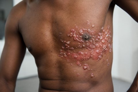
- Previous herpes zoster in the same area, neck or back injury or surgery, spinal symptoms, scar history.
- Diabetes, renal disease, thyroid disease, HIV, pregnancy.
- Household contacts with itch, swimming or hot tub use, beach exposure (cutaneous larva migrans), barefoot walking on sand or dusty floors (tungiasis).
- Previous topical corticosteroid or antifungal use, which alters the appearance of dermatophyte infection.
Examination
- Morphology: scale, an active edge with central clearing (tinea), sharply demarcated geometric patches (contact dermatitis), lichenified plaques on the nape, ankles, forearms or scrotum, violaceous flat topped papules at the wrists and ankles (lichen planus), erythematous scaly plaques over extensor surfaces (psoriasis), serpiginous raised tracks (cutaneous larva migrans), a white nodule with a central black dot on the foot (tungiasis), grouped vesicles in a dermatome (zoster).
- Wood lamp: coral red fluorescence in erythrasma.
- Neurological examination of the affected dermatome: light touch, pin prick and temperature sensation, power, reflexes, spinal tenderness and range of movement.
- Regional lymph nodes.
- Bedside tests: potassium hydroxide microscopy of skin scrapings, dermoscopy, skin swab.
Red flags
- Itch with limb weakness, sphincter disturbance or gait change (spinal cord or root compression).
- Facial itch or numbness with sensory loss or cranial nerve signs.
- Persistent unilateral eczema of the nipple or areola (Paget disease of the breast).
- A nonhealing ulcer, a changing pigmented lesion or an enlarging nodule.
- Patches or plaques unresponsive to treatment (cutaneous T cell lymphoma).
- Rapidly spreading erythema with fever (cellulitis).
- Itch with regional lymphadenopathy.
Special groups
- Children: atopic dermatitis, scabies, papular urticaria and tinea are most common. Use mild potency corticosteroids only on the face and flexures.
- Older adults: asteatotic eczema, transient acantholytic dermatosis and pemphigoid are more frequent. Avoid sedating antihistamines.
- Pregnancy: use emollients, hydrocortisone and moderate potency corticosteroids; avoid oral terbinafine and oral azoles.
- Region specific: tungiasis, cutaneous larva migrans, dermatophyte infections and onchocerciasis with nodules.
Inflammatory dermatoses
- Allergic contact dermatitis and irritant contact dermatitis.
- Atopic dermatitis and nummular eczema.
- Asteatotic eczema.
- Stasis dermatitis: lower legs, venous disease.
- Dyshidrotic eczema: hands and feet.
- Lichen simplex chronicus.
- Prurigo nodularis.
- Psoriasis.
- Lichen planus.
- Seborrhoeic dermatitis.
- Transient acantholytic dermatosis (Grover disease): trunk in older adults after heat or sweating.
Infection and infestation
- Tinea corporis, tinea pedis and tinea manuum.
- Cutaneous candidiasis and erythrasma.
- Scabies.
- Pediculosis corporis.
- Cutaneous larva migrans.
- Tungiasis.
- Insect bite reactions and papular urticaria.
- Herpes zoster prodrome and post herpetic itch.
- Impetigo and folliculitis.
- Pityriasis versicolor.
Neuropathic
- Notalgia paraesthetica.
- Brachioradial pruritus.
- Cervical or lumbar radiculopathy.
- Spinal cord lesion.
- Small fibre neuropathy.
- Scar or post burn itch.
- Trigeminal trophic syndrome.
- Central nervous system lesion (stroke, tumour, multiple sclerosis).
Neoplastic and premalignant
- Cutaneous T cell lymphoma (mycosis fungoides).
- Paget disease of the nipple and extramammary Paget disease.
- Bowen disease.
- Basal cell carcinoma and squamous cell carcinoma.
Other
- Macular and lichen amyloidosis.
- Cutaneous mastocytosis.
- Cholinergic urticaria.
- Polymorphic light eruption.
- Aquagenic pruritus.
- Neurotic excoriation.
- Diabetes mellitus and iron deficiency with localised itch.
Step 1. Morphology. Decide whether primary lesions are present.
Step 2. Lesions present
- Potassium hydroxide microscopy and fungal culture of scrapings from the active edge before any topical corticosteroid. False negatives are frequent after topical antifungal or corticosteroid use.
- Bacterial swab for weeping, crusted or pustular skin.
- Patch testing for suspected allergic contact dermatitis after 4 to 6 weeks without topical corticosteroid on the test site. Irritant reactions can mislead; interpret with exposure history.
- Skin punch biopsy (4 mm) for any persistent unexplained plaque, a suspected lymphoma (several biopsies, plus T cell receptor gene analysis), suspected Paget disease, amyloid or mastocytosis.
- Full blood count, ferritin, glucose or HbA1c, urea and creatinine, TSH, and HIV test when itch persists or lesions are atypical.
Step 3. Normal skin with dermatomal or regional itch
- Detailed neurological examination.
- MRI of the cervical spine for brachioradial pruritus with neurological signs or suspected cord or root disease. MRI of the thoracic spine for notalgia only if neurological deficit or red flags.
- Nerve conduction studies and skin biopsy for intraepidermal nerve fibre density when small fibre neuropathy is suspected.
- Systemic screen: full blood count, ferritin, glucose or HbA1c, urea and creatinine, TSH and HIV test.
Interpretation. A normal spine MRI does not exclude notalgia or brachioradial pruritus, which are often diagnosed clinically. Patch test positives need relevance to the patient's exposure.
General measures. Identify and remove triggers, emollients twice daily, cut nails, avoid rubbing, cooling with menthol 1% in aqueous cream. For lichen simplex chronicus, break the scratch cycle with occlusive dressings at night.
Topical corticosteroids. Apply a thin layer once daily. Use 1 fingertip unit (0.5 g) for an area equal to 2 adult palms.
- Face and flexures: hydrocortisone 1% once daily for up to 7 to 14 days.
- Trunk and limbs: mometasone furoate 0.1% once daily for 2 to 4 weeks.
- Lichenified plaques on limbs: clobetasol propionate 0.05% once daily for up to 2 weeks, maximum 50 g weekly. Not for the face or flexures.
- Monitor for skin atrophy, striae, perioral dermatitis and adrenal suppression with large areas. Do not apply to untreated infection.
Topical calcineurin inhibitors. Tacrolimus 0.1% ointment twice daily (0.03% for children) or pimecrolimus 1% cream twice daily on face and flexures. Burning at the start is common.
Cause directed
- Tinea corporis: terbinafine 1% cream once or twice daily for 1 to 2 weeks, or clotrimazole 1% cream 2 to 3 times daily for 2 to 4 weeks, continued 1 to 2 weeks after clearing. Extensive disease: terbinafine 250 mg orally once daily for 2 weeks (reduce to 125 mg daily if creatinine clearance is under 50 mL/min; avoid in active liver disease; not for pregnancy). Check liver tests if treatment exceeds 6 weeks.
- Scabies: permethrin 5% cream to the whole body from the neck down, left for 8 to 12 hours and repeated after 7 days, with simultaneous treatment of contacts. Ivermectin 200 micrograms/kg orally with food, repeated after 7 days, is an alternative (not for children under 15 kg or pregnancy).
- Cutaneous larva migrans: ivermectin 200 micrograms/kg orally as a single dose, or albendazole 400 mg orally once daily for 3 days.
- Tungiasis: sterile extraction of the flea, tetanus immunisation status check, topical dimeticone. Cover secondary bacterial infection with flucloxacillin 500 mg orally four times daily for 5 to 7 days (clarithromycin 250 to 500 mg twice daily if penicillin allergic).
- Impetigo and folliculitis: mupirocin 2% ointment three times daily for 5 days, or fusidic acid 2% cream three times daily for 5 to 7 days. Extensive disease: flucloxacillin 500 mg orally four times daily for 5 to 7 days (clarithromycin 250 to 500 mg twice daily if penicillin allergic).
- Neuropathic itch: capsaicin 0.025% to 0.075% cream applied 3 to 4 times daily for 2 to 6 weeks (initial burning is expected). Gabapentin 300 mg orally at night, titrated to 900 to 1800 mg daily in 3 divided doses, maximum 3600 mg daily, with renal dose reduction (creatinine clearance 30 to 59 mL/min, 400 to 1400 mg daily; 15 to 29 mL/min, 200 to 700 mg once daily; under 15 mL/min, 100 to 300 mg once daily). Sun protection and physiotherapy for brachioradial pruritus.
- Contact dermatitis: avoid the allergen and use hydrocortisone 1% or mometasone furoate 0.1% once daily for up to 7 days.
- Oral antihistamines help only urticarial or insect bite itch: cetirizine 10 mg orally once daily (5 mg daily if creatinine clearance is under 50 mL/min).
Monitoring. Review at 2 to 4 weeks. Reassess the diagnosis if there is no response. Check skin for atrophy.
Referral and escalation. Dermatology within 2 weeks for suspected skin cancer, lymphoma or Paget disease. Neurosurgical or spinal assessment for cord compression signs (same day). Dermatology for refractory prurigo nodularis (biologic therapy is available).
Disposition. Most patients are managed in primary care. Admit for cellulitis with systemic features or spinal cord compression. Safety net advice: return for new neurological symptoms, a nonhealing lesion, spreading redness with fever, or failure to improve after 4 weeks.
