



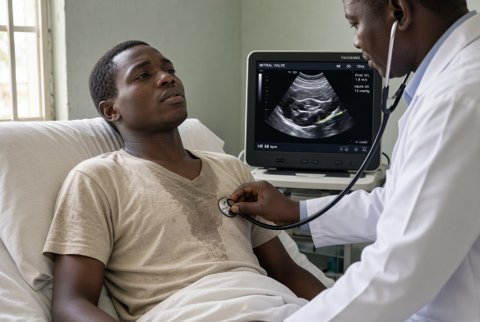








Postoperative abdominal distension
Postoperative abdominal distension is visible or measurable enlargement of the abdomen after surgery, from gas, fluid, bowel dilatation, blood, pus, urine or raised intra-abdominal pressure. Most cases are physiological ileus, but distension can signal obstruction, anastomotic leak, bleeding or abdominal compartment syndrome.
Clinical relevance. Postoperative ileus follows most abdominal operations and prolongs stay when it lasts over 4 to 5 days. Early small bowel obstruction accounts for a minority of cases and anastomotic leak carries a mortality of 10 to 20%.
Physiology. After laparotomy, the small bowel recovers motility within hours, the stomach within 24 to 48 hours and the colon within 3 to 5 days. Opioids, electrolyte disturbance, inflammation, sympathetic activation and handling of bowel slow this recovery.
Classification.
- Expected postoperative ileus: absent bowel sounds and flatus for up to 3 to 5 days, with tolerable distension.
- Prolonged postoperative ileus: persisting beyond 5 days after bowel surgery (or beyond 3 days after non-bowel surgery), or recurring after tolerating diet.
- Mechanical obstruction: adhesions, internal hernia, port site or incisional hernia, anastomotic stricture, volvulus, intussusception, retained foreign body.
- Colonic pseudo-obstruction (Ogilvie syndrome): colonic dilatation without a mechanical cause.
- Distension from collection or fluid: leak, abscess, haemoperitoneum, ascites, urine, chyle.
- Raised intra-abdominal pressure: intra-abdominal hypertension (sustained pressure of 12 mmHg or more) and abdominal compartment syndrome (over 20 mmHg with new organ dysfunction).
Key distinctions. Ileus is painless or mildly uncomfortable with generalised distension and absent bowel sounds. Obstruction causes colicky pain, vomiting and high pitched sounds. Leak causes fever, tachycardia, rising CRP and peritonism, usually from days 3 to 7.
History.
- Type, duration and urgency of the operation, bowel handling and anastomosis, drains, epidural use, fluid balance, and opioid dose.
- Day of onset, pattern (progressive or early), nausea, vomiting (bilious, faeculent), pain and its character, passage of flatus and stool, oral intake and tolerance of diet.
- Fever, cough, urinary retention, calf swelling, and abdominal wall pain or swelling.
- Drugs: opioids, anticholinergics, calcium channel blockers, antipsychotics, vincristine, clonidine, and recent changes.
- Prior abdominal surgery or hernia, radiotherapy, inflammatory bowel disease, constipation, malignancy and diabetes.
Examination.
- Vital signs, hydration, mental state, urine output and respiratory effort.
- Abdomen: degree and symmetry of distension, wound and port sites (hernia, collection, discolouration), tenderness, guarding, rebound, tympany or dullness, bowel sounds (absent in ileus, tinkling and high pitched in obstruction), and a succussion splash.
- Drains: volume and character (bile, enteric content, pus, blood). Stoma colour and output.
- Hernial orifices, rectal examination (impaction, blood, empty rectum), and bladder palpation.
- Calves, chest, and signs of sepsis.
Red flags.
- Fever, tachycardia over 100 beats/min, hypotension, rising lactate over 2 mmol/L, or oliguria (sepsis or leak).
- Peritonism, rigidity or severe pain out of proportion to the findings (perforation, ischaemia).
- Bilious or faeculent vomiting, or severe colicky pain with absent flatus (obstruction).
- Caecal diameter over 9 to 10 cm on imaging, or rapidly rising distension (risk of perforation, particularly over 12 cm).
- Bloody or enteric drain output, or falling haemoglobin (bleed or leak).
- Bladder pressure over 20 mmHg with oliguria, rising airway pressures or hypoxia (compartment syndrome).
- Irreducible tender hernia.
- Profuse diarrhoea with distension (severe Clostridioides difficile infection, toxic megacolon).
Special groups. Older adults and critically ill patients present with minimal pain and fever. Pregnancy after caesarean section: consider ileus, endometritis and bleeding. Children need weight based fluids and early surgical review.
Functional.
- Primary postoperative ileus, secondary ileus (sepsis, electrolyte disturbance, drug related, peritonitis, retroperitoneal haematoma, pancreatitis, pneumonia, myocardial infarction).
- Colonic pseudo-obstruction (Ogilvie syndrome), gastroparesis and delayed gastric emptying, constipation and faecal impaction.
Mechanical obstruction.
- Early adhesive small bowel obstruction, internal hernia (after bariatric and colorectal surgery), port site or incisional hernia, volvulus, intussusception.
- Anastomotic stricture or oedema, stoma obstruction, retained swab or foreign body, tumour recurrence, band.
Intra-abdominal complications. Anastomotic leak with peritonitis, intra-abdominal abscess, haemoperitoneum, bile leak, pancreatic fistula, chylous ascites, mesenteric ischaemia, bowel perforation, visceral injury, gossypiboma.
Raised pressure. Abdominal compartment syndrome, tense ascites, massive fluid resuscitation with bowel oedema, retroperitoneal haematoma.
Infective and inflammatory. Clostridioides difficile infection and toxic megacolon, pancreatitis, cholecystitis, appendicitis, intra-abdominal sepsis, necrotising infection.
Urinary and other. Urinary retention or bladder distension, ureteric injury with urinoma, hypoalbuminaemia with third spacing, cirrhosis with ascites, hypokalaemia, hypomagnesaemia, hypothyroidism, opioid effect.
Step 1. Assess for sepsis and peritonism immediately. Decide whether the distension is tolerated and improving (ileus) or progressing with systemic signs (leak, obstruction, ischaemia).
Step 2. First line tests: full blood count, CRP, urea, creatinine, electrolytes (potassium, magnesium, phosphate, calcium), glucose, lactate, liver tests, amylase or lipase, coagulation, blood gas, and group and save. Urinalysis and a bladder scan. Blood cultures if febrile.
Step 3. Imaging.
- Plain abdominal radiograph: gas pattern, dilated small bowel (over 3 cm) or colon (caecum over 6 cm, transverse colon over 5.5 cm), air fluid levels, free gas (expected for several days after laparotomy). Supine and erect or decubitus views.
- CT abdomen and pelvis with intravenous contrast (oral or rectal contrast depending on suspicion) is the best test for leak, collection, obstruction with a transition point, volvulus, ischaemia and pseudo-obstruction. A CT after day 5 to 7 is more reliable for leak.
- Water soluble contrast (diatrizoate 50 to 100 mL orally or by tube) with radiographs at 8 to 24 hours distinguishes partial from complete obstruction, and contrast reaching the colon within 24 hours predicts resolution.
- Ultrasound for collections, free fluid, hernias and bladder volume.
- Contrast enema for suspected colonic obstruction or pseudo-obstruction. Colonoscopy for decompression and diagnosis.
Step 4. Measure bladder pressure (via a urinary catheter, instilling 25 mL, supine at end expiration, zeroed at the mid axillary line) in at risk patients with tense distension, oliguria or high airway pressure. Grade I: 12 to 15 mmHg; II: 16 to 20; III: 21 to 25; IV: over 25 mmHg.
Interpretation. CRP over 150 mg/L on day 3 to 4 raises concern for leak or collection. Rising lactate, leucocytosis and a falling urine output after an initial recovery point to a complication. Drain fluid amylase, bilirubin, creatinine and culture can confirm leak.
Limitations. Plain films show non-specific dilatation. Contrast studies can miss small leaks. CT is hard to interpret in the first week after laparotomy.
Immediate priorities. Resuscitate (oxygen, IV access, isotonic crystalloid 20 mL/kg boluses for hypotension), stop oral intake, treat sepsis within 1 hour, and call the surgical team for peritonism, leak, ischaemia, strangulation or compartment syndrome.
Expected and prolonged ileus (supportive treatment).
- Early mobilisation, chewing gum, and early oral feeding when tolerated. Avoid fluid overload (target near zero balance after resuscitation) and treat pain with opioid sparing analgesia: paracetamol 1 g orally or IV every 6 hours (maximum 4 g daily; 3 g daily if weight under 50 kg, malnutrition, alcohol excess or liver disease), and regional or epidural analgesia when available.
- Nasogastric tube for vomiting, marked gastric distension or aspiration risk, left on free drainage, and removed when output is under 400 mL daily and the patient is hungry.
- Correct electrolytes: potassium to 4.0 mmol/L or more and magnesium to 0.8 mmol/L or more. For potassium of 3.0 to 3.5 mmol/L give 40 mmol potassium chloride in 1 L isotonic saline over 4 to 6 hours (maximum 10 mmol/hour peripherally, 20 mmol/hour centrally with ECG monitoring); for magnesium under 0.7 mmol/L give magnesium sulphate 2 g IV over 20 minutes to 2 hours (monitor reflexes and respiratory rate; reduce in kidney impairment).
- Stop or reduce opioids, anticholinergics and other drugs that slow motility. Laxatives and enemas for constipation once obstruction is excluded: macrogol 1 sachet orally once or twice daily, or senna 7.5 to 15 mg orally at night, or glycerol suppository 4 g rectally.
- Prokinetics have limited evidence: metoclopramide 10 mg IV or orally every 8 hours for up to 5 days (maximum 30 mg daily; 5 mg every 8 hours if CrCl under 40 mL/min; avoid in mechanical obstruction and in Parkinson disease; monitor for extrapyramidal effects), or erythromycin 250 mg IV every 8 hours for 3 to 5 days (monitor QTc and interactions).
- Nutrition: parenteral nutrition when enteral intake is not possible for 5 to 7 days, or sooner in malnourished patients; check refeeding risk with phosphate, magnesium and thiamine 100 to 300 mg IV daily.
Suspected mechanical small bowel obstruction. Nil by mouth, nasogastric decompression, intravenous isotonic crystalloid with potassium, and urgent surgical review. Adhesive obstruction without strangulation may resolve in 48 to 72 hours with water soluble contrast. Operate for peritonism, strangulation, closed loop, ischaemia, complete obstruction with no resolution at 48 to 72 hours, internal hernia, or an early postoperative obstruction that persists.
Colonic pseudo-obstruction.
- Exclude mechanical obstruction and ischaemia with CT. Treat the cause: stop opioids, anticholinergics and sedatives, correct electrolytes and treat sepsis. Nasogastric tube and a rectal tube, mobilise and position the patient, and monitor serial radiographs at 12 to 24 hours.
- Neostigmine 2 mg IV over 3 to 5 minutes (maximum 2.5 mg; repeat once after 3 hours when partially effective) when caecal diameter is over 10 cm or the patient is not improving after 24 to 48 hours of supportive treatment, with continuous ECG and blood pressure monitoring and atropine 0.6 to 1.2 mg IV immediately available; avoid with mechanical obstruction, bradycardia under 60 beats/min, bronchospasm, recent myocardial infarction, active ischaemia, pregnancy and kidney impairment with CrCl under 30 mL/min (reduce the dose to 1 mg). Expect abdominal cramping, bradycardia, salivation and bronchospasm.
- Colonoscopic decompression when neostigmine fails or is contraindicated, caecal or percutaneous decompression for perforation risk, and surgery for perforation or ischaemia.
Anastomotic leak or intra-abdominal collection.
- Sepsis resuscitation, nil by mouth, and urgent source control: percutaneous drainage of a contained collection, or laparotomy for generalised peritonitis, uncontained leak or instability.
- Piperacillin with tazobactam 4.5 g IV every 6 hours (maximum 18 g piperacillin daily; every 8 hours for CrCl 20 to 40 mL/min and every 12 hours under 20 mL/min; monitor creatinine and full blood count), or meropenem 1 g IV every 8 hours (1 g every 12 hours for CrCl 26 to 50 mL/min, 500 mg every 12 hours for CrCl 10 to 25 mL/min, 500 mg every 24 hours under 10 mL/min; monitor seizures and creatinine) when resistant organisms are likely.
- Severe penicillin allergy: ciprofloxacin 400 mg IV every 12 hours (maximum 1.2 g daily) plus metronidazole 500 mg IV every 8 hours (maximum 1.5 g daily); monitor QTc, tendon pain and glucose; reduce the dose in kidney impairment.
- Review at 48 to 72 hours with cultures, and continue for about 4 to 7 days after adequate source control.
Abdominal compartment syndrome. Decompress (nasogastric and rectal tubes, prokinetics), drain tense ascites or collections, remove constrictive dressings, optimise sedation and analgesia, consider neuromuscular blockade under critical care direction, avoid fluid overload, and proceed to decompressive laparotomy for pressure over 20 mmHg with organ failure that does not respond. Admit to critical care.
Haemoperitoneum. Resuscitate with blood products, correct coagulopathy, and return to theatre or embolisation for ongoing bleeding.
Urinary retention. Catheterise (record volume), review drugs, and remove the catheter after 24 to 48 hours when the patient is mobile and passing urine.
Clostridioides difficile infection. Vancomycin 125 mg orally every 6 hours for 10 days (maximum 500 mg daily), or 500 mg every 6 hours orally or by nasogastric tube plus metronidazole 500 mg IV every 8 hours for fulminant disease with ileus (maximum vancomycin 2 g daily); urgent surgical review for toxic megacolon.
Monitoring. Abdominal girth, vital signs every 4 hours (hourly in sepsis), fluid balance, nasogastric output, bowel function and flatus, daily electrolytes, repeat imaging when the clinical picture changes, and bladder pressure every 4 to 6 hours for intra-abdominal hypertension.
Referral and escalation. Senior surgeon for any suspected complication. Critical care for sepsis, shock, compartment syndrome and pseudo-obstruction needing monitored neostigmine. Gastroenterology for colonoscopic decompression. Interventional radiology for drainage and embolisation. Dietitian for prolonged ileus.
Admission criteria. All inpatients with distension and vomiting, sepsis, peritonism, caecal diameter over 10 cm, suspected obstruction, leak or compartment syndrome. Patients discharged recently who return with distension, vomiting, fever or pain are readmitted.
Discharge and safety netting. Discharge when eating, passing flatus, afebrile, and pain controlled on oral analgesia. Patients return for vomiting, increasing distension, absent flatus, abdominal pain, fever, wound discharge, or reduced urine output.
