
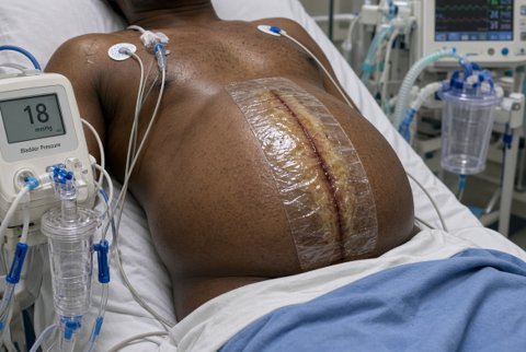










Abdomen Overview
Superior: Diaphragm, separating the abdominal cavity from the thoracic cavity (dome extends as high as the 4th-5th intercostal space anteriorly, meaning upper abdominal organs (liver, spleen, stomach) are partly protected by the lower rib cage).
- Inferior: Pelvic inlet, continuous with the pelvic cavity (no true anatomical barrier, the abdominal and pelvic cavities are functionally continuous).
- Anterior/lateral: Anterior abdominal wall muscles (external oblique, internal oblique, transversus abdominis, rectus abdominis).
- Posterior: Vertebral column (lumbar vertebrae) and posterior abdominal wall muscles (psoas major, quadratus lumborum, iliacus).
- Musculofascial envelope surrounding the abdominal cavity, providing structural support, protection, and contributing to intra-abdominal pressure generation.
- Anterolateral wall: three flat muscle layers plus rectus abdominis; posterior wall, psoas, quadratus lumborum, iliacus, overlying the vertebral column.
- The space bounded by the abdominal wall, diaphragm, and pelvic inlet, containing the abdominal viscera, peritoneum, and retroperitoneal structures.
- Often considered together with the pelvic cavity as the abdominopelvic cavity, given their continuity.
- Potential space within the abdominal cavity, between the parietal and visceral peritoneum, containing a thin film of serous fluid.
- Subdivided into the greater sac (the main peritoneal cavity) and the lesser sac (omental bursa), connected via the epiploic foramen; full detail in the Peritoneum section.
- Foregut derivatives: Stomach, first/second parts of the duodenum, liver, gallbladder, pancreas, spleen (though the spleen is not a true gut derivative, it shares the foregut's vascular supply via the coeliac trunk).
- Midgut derivatives: Distal duodenum, jejunum, ileum, caecum, appendix, ascending colon, proximal two-thirds of the transverse colon.
- Hindgut derivatives: Distal third of the transverse colon, descending colon, sigmoid colon, rectum (upper part).
- Retroperitoneal viscera: Kidneys, ureters, suprarenal glands, and (secondarily retroperitoneal) the duodenum (parts 2-4), pancreas (most of it), ascending and descending colon.
- Structures lying posterior to the parietal peritoneum, against the posterior abdominal wall.
- Primary retroperitoneal (never had a mesentery, developed outside the peritoneum), kidneys, ureters, suprarenal glands, abdominal aorta, IVC.
- Secondary retroperitoneal (originally intraperitoneal with a mesentery, later fused to the posterior wall during development), duodenum (2nd-4th parts), pancreas (head, neck, body), ascending colon, descending colon.
- Clinically relevant to surgical access (retroperitoneal structures can sometimes be approached without entering the peritoneal cavity) and to the pattern of retroperitoneal haemorrhage/pathology spread.
- Abdominal aorta: descends from the aortic hiatus (T12) to its bifurcation (L4), giving unpaired visceral branches (coeliac trunk, SMA, IMA), paired visceral branches (renal, gonadal, middle suprarenal), and paired parietal branches (inferior phrenic, lumbar).
- Inferior vena cava: formed by the union of the common iliac veins (L5), ascends to the diaphragm's caval opening (T8), draining the lower limbs, pelvis, and abdominal viscera (except the portal-drained gut, which passes through the liver first).
- Portal vein: formed by the union of the splenic and superior mesenteric veins, carries nutrient-rich venous blood from the gut to the liver.
- Lumbar plexus (T12-L4), iliohypogastric, ilioinguinal, genitofemoral, lateral femoral cutaneous, femoral, obturator nerves.
- Sympathetic trunks and splanchnic nerves: continue from the thorax, synapsing in prevertebral ganglia around the aortic branch origins.
- Vagus nerve: parasympathetic supply to foregut and midgut structures, continuing from the thoracic oesophageal plexus.
- Four-quadrant system (simpler, commonly used clinically): right upper quadrant (RUQ), left upper quadrant (LUQ), right lower quadrant (RLQ), left lower quadrant (LLQ), divided by a vertical (midline) and horizontal (through the umbilicus) plane.
- Nine-region system (more detailed, used for precise anatomical description): right/left hypochondrium, epigastric, right/left lumbar (flank), umbilical, right/left iliac (inguinal), hypogastric (suprapubic), divided by two vertical (midclavicular) lines and two horizontal planes (subcostal and transtubercular, or historically transpyloric and intertubercular).
- Transpyloric plane (of Addison): Horizontal plane midway between the jugular notch and pubic symphysis, approximately at the L1 vertebral level: a critical reference plane, correlating with the pylorus of the stomach, the neck of the pancreas, the hila of the kidneys, the origin of the SMA, the formation of the portal vein, the termination of the spinal cord (approximately), and the fundus of the gallbladder (at its intersection with the right costal margin/midclavicular line).
- Subcostal plane: Horizontal plane connecting the lowest points of the costal margins (10th costal cartilage), approximately at the L3 level.
- Transtubercular plane: Horizontal plane connecting the iliac tubercles, approximately at the L5 level: used with the midclavicular lines to define the nine-region grid.
- Intercristal (supracristal) plane: Connects the highest points of the iliac crests, approximately at the L4 level: a key landmark for safe lumbar puncture (below the level of the spinal cord's termination).
- The gastrointestinal tract is organised embryologically and vascularly into foregut (coeliac trunk), midgut (SMA), and hindgut (IMA) territories, a unifying framework explaining shared blood supply, innervation, and referred pain patterns across seemingly separate organs.
- The abdomen functions as the primary site of digestion, absorption, and metabolic processing (liver), alongside filtration/excretion (kidneys) and additional endocrine/immune functions (pancreas, spleen, adrenals).
