









Fungal Stains and Culture
Fungal stains and culture detect yeasts, moulds and dimorphic fungi from skin, hair, nails, mucosa, respiratory samples, blood, CSF, sterile fluids and tissue.
Direct microscopy is rapid, with potassium hydroxide (KOH) 10 to 20% clearing keratin and tissue, calcofluor white or Blankophor fluorescence (the most sensitive), lactophenol cotton blue, Gram stain for yeasts, Grocott-Gomori methenamine silver, periodic acid-Schiff, India ink for Cryptococcus and Giemsa for Histoplasma and Talaromyces.
Culture is on Sabouraud dextrose agar (with chloramphenicol and with or without cycloheximide), brain heart infusion agar, chromogenic Candida agar, and special media (dermatophyte test medium, birdseed agar), incubated at 25 to 30 °C and 35 to 37 °C for 2 to 4 weeks (up to 6 weeks for dimorphic fungi).
Blood culture systems detect Candida, but are poor for moulds and Histoplasma (lysis centrifugation is better). Identification uses morphology, MALDI-TOF mass spectrometry and DNA sequencing, followed by antifungal susceptibility testing.
Fungal infections affect over a billion people, with superficial infections (tinea, candidiasis) the commonest, and invasive infections (candidaemia, invasive aspergillosis, cryptococcosis, histoplasmosis, mucormycosis, Pneumocystis pneumonia, talaromycosis) causing about 1.5 million deaths each year.
Invasive disease concentrates in immunocompromised patients (haematological malignancy, transplantation, prolonged neutropenia, corticosteroids, HIV, intensive care, diabetic ketoacidosis, broad spectrum antibiotics, central venous catheters and total parenteral nutrition).
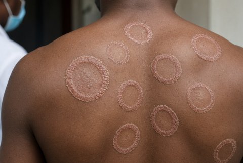
Indications. Suspected dermatophyte infection of skin, hair or nails (before oral therapy, since treatment is prolonged and toxic). Oral and oesophageal candidiasis not responding to treatment. Fever in a neutropenic patient unresponsive to antibiotics. Pulmonary nodules, cavities or infiltrates in an immunocompromised patient.
Sinusitis with black necrotic tissue or palate lesions (mucormycosis). Fungal keratitis, endophthalmitis, sporotrichosis, mycetoma and chromoblastomycosis. Chronic meningitis, osteomyelitis or lymphadenitis. Endemic mycoses after exposure (histoplasmosis, coccidioidomycosis, blastomycosis, talaromycosis).
Specimen. Skin: scrape scales from the active edge of the lesion after cleaning with 70% alcohol. Hair: plucked hairs with roots. Nails: clippings and subungual debris from the deep proximal portion. Mucosal swabs.
Sputum, bronchoalveolar lavage, tissue biopsy (the best specimen for invasive disease, sent in a sterile container with saline and not in formalin for culture), aspirates, CSF, blood (at least two sets, with an adequate volume), and urine. Transport promptly. Never refrigerate specimens for dimorphic fungi or Mucorales, and do not grind tissue, which damages the hyphae of Mucorales.
