








Haemoptysis
Haemoptysis is the coughing up of blood from the lower respiratory tract (larynx, trachea, bronchi or lung parenchyma). It ranges from blood streaking of sputum to massive bleeding that threatens life by asphyxiation rather than by blood loss. The first decisions are whether the blood truly came from the airway (not the mouth, nose or stomach), how much is being lost, and whether the airway and oxygenation are secure.
Clinical relevance. Most episodes are mild and self limiting, and an apparent cause is found in about 70% to 80%: bronchitis and bronchiectasis, pneumonia, tuberculosis and lung cancer lead in adults. About 20% to 30% are cryptogenic after full work up. Massive haemoptysis is uncommon (under 5%) but carries mortality of 10% to 50% without control, since death is from airway flooding. Lung cancer must be excluded in every smoker over 40 with haemoptysis.
Classification.
- By volume: scant or streaking (under 5 mL in 24 hours), mild to moderate (5 to 100 mL), severe (over 100 mL in 24 hours), massive (over 200 to 600 mL in 24 hours, or any volume that compromises gas exchange or airway). Rate matters more than total: 100 mL in an abnormal lung can be lethal.
- By source: bronchial arterial (systemic, high pressure, 90% of massive bleeding), pulmonary arterial (low pressure, for example cavity erosion, Rasmussen aneurysm, arteriovenous malformation), capillary or diffuse alveolar (vasculitis, anti glomerular basement membrane disease, mitral stenosis).
- By cause: airway, parenchymal, vascular, haematological and drug related, traumatic and iatrogenic, cryptogenic.
- Pseudohaemoptysis: blood from nose, throat, gums or upper gastrointestinal tract (haematemesis), or red coloured sputum from Serratia marcescens or drugs (rifampicin).
Mechanism. Chronic inflammation causes hypertrophy and tortuosity of bronchial arteries and anastomoses with pulmonary vessels, which rupture under systemic pressure. Tumour, infection, cavities, necrosis, vasculitis and raised pulmonary venous pressure all injure vessels and alveolar capillaries.
History.
- Quantity and colour (bright red, frothy suggests airway origin; dark, coffee ground with food suggests gastric), timing, number of episodes, and whether preceded by cough and a tickle (airway) or nausea and vomiting (stomach).
- Nose bleed, dental or throat symptoms, recent facial or oral procedure.
- Cough duration, sputum character, fever, night sweats, weight loss, chest pain (pleuritic suggests embolism or pneumonia), breathlessness, wheeze, hoarseness.
- Smoking (pack years), cannabis or crack cocaine use, occupational exposure (asbestos, silica), previous tuberculosis or contacts, residence or travel in endemic regions (tuberculosis, paragonimiasis, hydatid disease, melioidosis, aspergillosis), HIV risk.
- Leg swelling, recent surgery, immobility, previous thrombosis, oestrogen use, cancer (pulmonary embolism).
- Orthopnoea, palpitations, rheumatic fever (mitral stenosis, heart failure).
- Haematuria, rash, joint pain, sinusitis, nasal crusting (vasculitis, Goodpasture disease), recurrent nose bleeds or family history of bleeding (hereditary haemorrhagic telangiectasia).
- Drugs: anticoagulants, antiplatelets, thrombolytics, NSAIDs, amiodarone, nitrofurantoin, cocaine, bevacizumab, other targeted cancer agents.
- Past lung disease: bronchiectasis, cystic fibrosis, chronic obstructive pulmonary disease, aspergilloma, sarcoidosis, lung biopsy or bronchoscopy, thoracic trauma, pulmonary hypertension.
Examination.
- Airway, breathing and circulation: respiratory rate, saturation, work of breathing, pulse, blood pressure, perfusion, mental state, ability to speak.
- Source: inspect the nose, mouth, tongue, pharynx and gums; telangiectasia of lips and tongue.
- Chest: localised wheeze (endobronchial lesion), crackles, bronchial breathing, pleural rub, dullness (effusion, collapse), signs of mitral stenosis (loud S1, opening snap, mid diastolic murmur), right ventricular heave.
- Systemic: clubbing (cancer, bronchiectasis, abscess), cachexia, lymph nodes (supraclavicular), hepatomegaly, calf swelling or tenderness, rash, purpura, nasal septal perforation, saddle nose, ear disease, vasculitic lesions, joint swelling.
- Fever, signs of endocarditis (new murmur, splinters), pulsatile masses (aneurysm).
Red flags.
- Haemoptysis over 100 mL in 24 hours, or any ongoing bleeding with breathlessness or desaturation (massive haemoptysis).
- Saturation under 92%, stridor, inability to clear the airway, asphyxia.
- Haemodynamic instability.
- Smoker or ex smoker over 40 (lung cancer), unexplained weight loss, hoarseness, clubbing, supraclavicular nodes.
- Fever, night sweats, tuberculosis exposure, HIV (tuberculosis).
- Pleuritic chest pain, leg swelling, recent surgery (pulmonary embolism).
- Haematuria with haemoptysis (pulmonary renal syndrome).
- Anticoagulation or coagulopathy with bleeding.
- Previous lung cavity or aspergilloma with recurrent bleeding.
- Injecting drug use with fever (septic emboli).
Airway. Acute bronchitis, chronic bronchitis, bronchiectasis (including cystic fibrosis), lung cancer (squamous, small cell, adenocarcinoma), carcinoid and other endobronchial tumours, metastases, foreign body, bronchial trauma, Dieulafoy lesion of the bronchus, bronchovascular fistula, tracheo innominate fistula, broncholithiasis. Parenchymal infection.
Pneumonia (Klebsiella, Staphylococcus aureus, pneumococcus, Legionella), lung abscess, necrotising pneumonia, tuberculosis (active and post tuberculous cavity, Rasmussen aneurysm), nontuberculous mycobacteria, aspergilloma and chronic pulmonary aspergillosis, invasive fungal disease, paragonimiasis, hydatid cyst, melioidosis.
Vascular. Pulmonary embolism with infarction, pulmonary arteriovenous malformation (hereditary haemorrhagic telangiectasia), aortic aneurysm with bronchial fistula, pulmonary hypertension, bronchial artery aneurysm, mitral stenosis, left heart failure and pulmonary oedema, Eisenmenger syndrome, congenital heart disease with systemic to pulmonary collaterals.
Diffuse alveolar haemorrhage and immune. Granulomatosis with polyangiitis, microscopic polyangiitis, anti glomerular basement membrane disease, systemic lupus erythematosus, antiphospholipid syndrome, Behcet disease, idiopathic pulmonary haemosiderosis, eosinophilic granulomatosis with polyangiitis.
Haematological and drug related. Anticoagulants, thrombolysis, thrombocytopenia, disseminated intravascular coagulation, haemophilia and von Willebrand disease, platelet dysfunction, cocaine, amphetamine lung injury, amiodarone toxicity, bevacizumab and other antiangiogenic agents, penicillamine.
Traumatic and iatrogenic. Chest trauma (pulmonary contusion, bronchial rupture), lung biopsy, bronchoscopy, pulmonary artery catheter rupture, tracheostomy erosion, radiotherapy.
Pseudohaemoptysis. Epistaxis, gum disease, pharyngeal and laryngeal lesions, haematemesis, red pigmented sputum.
Unknown. Cryptogenic haemoptysis (up to 30%).
Clue | Think of |
|---|---|
Smoker over 40, weight loss, clubbing, persistent cough | Lung cancer |
Daily purulent sputum, recurrent infections, blood streaking | Bronchiectasis |
Fever, night sweats, weight loss, upper lobe cavity | Pulmonary tuberculosis |
Previous cavity, sudden large bright blood | Aspergilloma or post tuberculous cavity |
Pleuritic pain, calf swelling, tachycardia, hypoxia | Pulmonary embolism with infarction |
Orthopnoea, pink frothy sputum, mitral murmur | Mitral stenosis or pulmonary oedema |
Haematuria, nasal crusting, sinusitis, rash | Granulomatosis with polyangiitis or vasculitis |
Haematuria, rapidly progressive renal failure | Anti glomerular basement membrane disease |
Recurrent nose bleeds, lip telangiectasia, family history | Hereditary haemorrhagic telangiectasia |
Fever, productive cough, focal consolidation, rust coloured sputum | Pneumonia |
Injecting drug use, fever, multiple nodules | Septic emboli |
Haemoptysis on anticoagulant with high INR | Anticoagulant related bleeding |
Blood preceded by nausea, with food, dark | Haematemesis |
- Assess airway, breathing and circulation first; classify severity (massive if over 200 mL in 24 hours or compromised gas exchange).
- Confirm source: examine nose, mouth and throat; nasendoscopy if upper airway source is possible.
- Chest radiograph (posteroanterior and lateral) in all: mass, cavity, consolidation, infiltrates, bronchiectasis, nodules, effusion, collapse, pulmonary congestion; a normal radiograph does not exclude cancer or bronchiectasis.
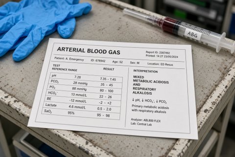
- Blood tests: full blood count (anaemia, platelets), coagulation (INR, activated partial thromboplastin time, fibrinogen), group and screen (crossmatch if severe), urea, creatinine, electrolytes, liver function, C reactive protein, arterial blood gas, lactate, urinalysis for blood and protein with urine microscopy (red cell casts), HIV test.
- Targeted tests: D-dimer and CT pulmonary angiography; antineutrophil cytoplasmic antibody, anti glomerular basement membrane antibody, antinuclear antibody, complement, antiphospholipid antibodies, anti DNA; sputum smear, culture, nucleic acid amplification for tuberculosis (3 samples), fungal culture, Aspergillus precipitins or IgG, cytology; blood cultures; interferon gamma release assay; natriuretic peptide and echocardiography.
- CT chest with contrast and CT angiography of the bronchial and systemic arteries (preferably before bronchoscopy; detects source, bronchiectasis, cancer, cavity, aneurysm, malformation, and guides embolisation); high resolution CT for diffuse disease.
- Bronchoscopy: in stable patients with an unexplained cause or risk of cancer (age over 40, smoker, abnormal radiograph) to find the bleeding site, take biopsies, washings, and cytology; rigid bronchoscopy for massive bleeding and removal of clots; flexible bronchoscopy for localisation and tamponade; bronchoalveolar lavage for diffuse alveolar haemorrhage (progressively bloodier return in successive aliquots).
- Lung biopsy, kidney biopsy and specialist work up for suspected vasculitis; cardiac investigation for suspected valve disease.
- Cryptogenic haemoptysis: CT and bronchoscopy negative; follow up for 6 to 12 months and repeat imaging.
Limits. Radiograph misses a source in up to 30%. CT detects bronchiectasis and small tumours better than bronchoscopy for peripheral lesions, while bronchoscopy sees endobronchial disease and is therapeutic. Bleeding site may be hard to lateralise. Diffuse alveolar haemorrhage can occur without haemoptysis. A normal chest radiograph and CT with a negative bronchoscopy still leaves a small risk of cancer, so smokers need follow up.
Massive haemoptysis (emergency).
- Call senior help, critical care, respiratory, interventional radiology and thoracic surgery at once.
- Airway first: position the patient bleeding side down (lateral decubitus) when the side is known, to protect the good lung; high flow oxygen; suction; intubate for exhaustion or hypoxia (use a large tube, 8.0 mm, to allow bronchoscopy); selective intubation of the non bleeding main bronchus or a double lumen tube or bronchial blocker when bleeding is torrential.
- Two large bore intravenous cannulas, group and crossmatch, transfuse red cells to keep haemoglobin over 7 to 8 g/dL (higher if shocked), correct coagulopathy (fresh frozen plasma, platelets to over 50 x 10^9/L, vitamin K 10 mg intravenously, prothrombin complex concentrate for warfarin; reversal agent for direct oral anticoagulants: idarucizumab 5 g for dabigatran, andexanet alfa for rivaroxaban and apixaban).
- Tranexamic acid: 1 g intravenously over 10 minutes then 1 g over 8 hours (reduces bleeding duration); or nebulised tranexamic acid 500 mg (5 mL of 100 mg/mL) three times daily for up to 5 days.
- Cough suppression and sedation only with ventilation protection (codeine 30 mg or morphine 2.5 mg intravenously for anxiety and cough in non ventilated patients with care).
- Bronchial artery embolisation (first line; control in 70% to 90% immediately, recurrence in 10% to 30%); needs CT angiography to identify non bronchial systemic arteries; avoid anterior spinal artery supply.
- Bronchoscopic control: cold saline lavage (4 degrees Celsius, 50 mL aliquots), topical adrenaline 1:10,000 (1 to 2 mL aliquots up to 10 mL), balloon tamponade, endobronchial blockers, laser or argon plasma coagulation.
- Surgical resection (lobectomy) for localised life threatening bleeding that fails embolisation (aspergilloma, abscess, cavity, tumour), in fit patients.
- Treat the cause: stop anticoagulants (or reverse), antibiotics, antituberculous therapy, vasculitis therapy.
Non massive haemoptysis.
- Reassure; rest; head of bed raised; sit upright or lie on the side of the bleed.
- Stop NSAIDs and antiplatelets if safe; hold anticoagulants and replace or reverse where the bleeding is significant and the indication allows (reassess the indication).
- Tranexamic acid 1 g orally or intravenously three times daily for 3 to 5 days (or nebulised 500 mg three times daily) for mild to moderate bleeding.
- Cough suppressant only if cough is exhausting (codeine linctus 15 mg three times daily); avoid sedatives that suppress cough in ventilated patients.
- Antibiotics for infection: community acquired pneumonia (amoxicillin 500 mg to 1 g three times daily for 5 to 7 days; co-amoxiclav 625 mg three times daily; doxycycline 200 mg then 100 mg daily), exacerbation of bronchiectasis (amoxicillin 500 mg to 1 g three times daily or doxycycline for 14 days; ciprofloxacin 500 to 750 mg twice daily for Pseudomonas), lung abscess (co-amoxiclav 1.2 g every 8 hours intravenously, or clindamycin 600 mg every 6 hours, then oral for 4 to 6 weeks).
- Tuberculosis: isoniazid 5 mg/kg to a maximum of 300 mg, rifampicin 10 mg/kg to a maximum of 600 mg, pyrazinamide 25 mg/kg to a maximum of 2 g, ethambutol 15 mg/kg for 2 months, then isoniazid and rifampicin for 4 months, with pyridoxine 10 mg daily; isolate; notify; contact tracing.
- Pulmonary embolism with haemoptysis is not a contraindication to anticoagulation: rivaroxaban 15 mg twice daily for 21 days then 20 mg daily, or apixaban 10 mg twice daily for 7 days then 5 mg twice daily, or enoxaparin 1 mg/kg twice daily, with close observation.
- Aspergilloma: bronchial artery embolisation, surgical resection if fit, itraconazole 200 mg twice daily or voriconazole 400 mg twice daily on day 1 then 200 mg twice daily for chronic pulmonary aspergillosis (monitor levels).
- Diffuse alveolar haemorrhage and vasculitis: methylprednisolone 500 to 1,000 mg intravenously daily for 3 days then prednisolone 1 mg/kg daily, plus rituximab 375 mg/m2 weekly for 4 weeks (or 1 g twice 2 weeks apart) or cyclophosphamide 15 mg/kg intravenously every 2 to 3 weeks (adjust for age and renal function), plasma exchange for anti glomerular basement membrane disease or severe renal involvement, supportive ventilation; specialist care.
- Heart failure and mitral stenosis: diuretics (furosemide 40 mg intravenously), rate control, balloon valvotomy or valve surgery.
- Hereditary haemorrhagic telangiectasia: embolisation of pulmonary arteriovenous malformations (feeding artery 3 mm or more), antibiotic prophylaxis before dental procedures, air filters on intravenous lines.
- Lung cancer: urgent staging and multidisciplinary treatment; palliative radiotherapy or endobronchial treatment for bleeding.
- Cryptogenic: observe; treat bronchitis; stop smoking.
- Stop smoking and avoid cocaine.
Disposition.
- Admit: any haemoptysis over 5 mL in a day that is not clearly trivial; recurrent bleeding; hypoxia; unstable vital signs; anticoagulation or coagulopathy; suspected cancer, tuberculosis, vasculitis or embolism; comorbid lung disease; social concerns.
- Intensive care or high dependency: massive haemoptysis, respiratory compromise, need for airway protection, embolisation.
- Home: streaking only in a young non smoker with a normal examination, normal chest radiograph and reliable follow up (review within 1 week), with urgent outpatient CT and bronchoscopy as indicated.
- Urgent referral (2 week suspected cancer) for smokers over 40, abnormal radiograph or persistent haemoptysis.
Monitoring. Oxygen saturation, respiratory rate, volume of blood loss, haemoglobin, coagulation, chest radiograph; observe closely for 24 to 48 hours after embolisation; repeat CT or bronchoscopy for persisting bleeding.
Referral and escalation. Respiratory physician for all haemoptysis needing investigation; interventional radiology for embolisation; thoracic surgery for resection; infectious diseases for tuberculosis and fungal disease; rheumatology and nephrology for vasculitis; haematology for coagulopathy; oncology.
Safety netting. Return urgently for coughing more than a teaspoon of blood, repeated bleeding, breathlessness, chest pain, fever, dizziness or collapse. Stop smoking and avoid NSAIDs and aspirin unless prescribed.
Special groups. Children: foreign body, bronchiectasis, congenital heart disease, tuberculosis, cystic fibrosis and bleeding disorders; embolisation is possible but technically hard. Pregnancy: mitral stenosis, pulmonary embolism, tuberculosis, pulmonary arteriovenous malformations (risk of rupture in pregnancy); avoid radiation where possible but do not delay essential CT. Older adults: cancer, aspergilloma, anticoagulation and aspiration.
Immunosuppressed and HIV: tuberculosis, fungal infection and Kaposi sarcoma. Cystic fibrosis: bleeding of 240 mL or more needs urgent embolisation; stop nonsteroidal drugs and hold intravenous antibiotics that affect clotting as needed. Endemic regions: tuberculosis, paragonimiasis, hydatid disease, schistosomiasis and melioidosis must be considered.
