








Pleuritic chest pain
Pleuritic chest pain is sharp, stabbing pain that is worse on inspiration, coughing, sneezing or movement and is relieved by shallow breathing or holding the breath. It arises from the parietal pleura (innervated by intercostal nerves), the pericardium, the diaphragm, the chest wall or mediastinal structures. The pain is often unilateral and may refer to the shoulder (diaphragmatic pleura via the phrenic nerve) or the abdomen.
Clinical relevance. The principal causes are pulmonary embolism, pneumonia, pneumothorax and pericarditis, but musculoskeletal, viral and autoimmune causes are frequent. Pulmonary embolism presents with pleuritic pain in about 60% of cases. Pleuritic pain from pneumonia may precede cough and fever.
Classification by origin.
- Pleural: pleurisy from infection (pneumonia, tuberculosis, viral), pulmonary infarction, malignancy, autoimmune disease (lupus, rheumatoid arthritis), asbestos related disease.
- Pericardial: pericarditis (pain is also postural and relieved by sitting forward).
- Vascular: pulmonary embolism with infarction, aortic dissection (usually not pleuritic).
- Airway: pneumothorax, pneumomediastinum.
- Chest wall: rib fracture, costochondritis, intercostal muscle strain, herpes zoster, epidemic pleurodynia (Coxsackie B).
- Subdiaphragmatic: hepatic and splenic disease, subphrenic abscess, pancreatitis, cholecystitis, perforated viscus.
History.
- Onset (sudden suggests embolism or pneumothorax; days suggests infection), side, site, radiation (shoulder tip pain), severity, relation to breathing, coughing, position and movement.
- Associated: breathlessness, cough (sputum colour, blood), fever, rigors, night sweats, weight loss, leg swelling or pain, palpitations, haemoptysis, rash, joint pain.
- Risk factors for embolism: immobilisation, surgery, trauma, cancer, previous thromboembolism, oestrogen, pregnancy, thrombophilia, long travel.
- Risk for pneumothorax: tall thin male, smoking, chronic lung disease, Marfan syndrome, recent procedures, mechanical ventilation.
- Infection: recent respiratory illness, tuberculosis contact, HIV, travel, aspiration risk, intravenous drug use.
- Autoimmune features: rash, mouth ulcers, joint pain, photosensitivity.
- Drug: hydralazine, procainamide, isoniazid, methotrexate, amiodarone, nitrofurantoin (drug induced lupus or pleuritis).
- Trauma, coughing bouts, rib tenderness, manual labour.
Examination.
- Vital signs, oxygen saturation, respiratory rate, temperature.
- Chest: splinting, shallow breathing, reduced expansion on the affected side, pleural rub, reduced breath sounds, dullness (effusion), hyperresonance (pneumothorax), bronchial breathing and crackles (consolidation).
- Cardiac: pericardial rub (scratchy, three components), tachycardia, raised jugular venous pressure, loud pulmonary second sound.
- Legs: swelling, calf tenderness, differential circumference over 3 cm.
- Chest wall: focal rib tenderness, vesicular rash, costochondral tenderness.
- Skin and joints (lupus rash, arthritis), lymph nodes, clubbing.
- Abdomen: right upper quadrant tenderness, epigastric tenderness.
Red flags.
- Hypoxia, respiratory rate over 24, heart rate over 110, hypotension, syncope.
- Haemoptysis; unilateral leg swelling; known cancer.
- Tracheal deviation, hyperresonance with distress (tension pneumothorax).
- Large effusion with dyspnoea, empyema (fever, effusion).
- Pericardial effusion with tamponade features.
- Severe sepsis with pneumonia.
- Immunosuppression, HIV, sickle cell disease (acute chest syndrome).
Thromboembolic and vascular. Pulmonary embolism with infarction, septic emboli (endocarditis, intravenous drug use), sickle cell acute chest syndrome, vasculitis (granulomatosis with polyangiitis).
Infective. Community acquired pneumonia (pneumococcal, atypical), tuberculosis (pleurisy with effusion), viral pleurisy, parapneumonic effusion and empyema, lung abscess, Coxsackie B pleurodynia, fungal infection, hydatid disease, amoebic liver abscess, subphrenic abscess.
Pleural and airway. Primary and secondary spontaneous pneumothorax, traumatic pneumothorax, pneumomediastinum, malignant pleural effusion, mesothelioma, lung cancer, haemothorax, chylothorax, asbestos pleural disease.
Pericardial. Acute pericarditis (viral, tuberculous, uraemic, post infarction, autoimmune, malignant), pericardial effusion.
Autoimmune. Systemic lupus erythematosus, rheumatoid arthritis, familial Mediterranean fever, drug induced lupus, sarcoidosis, adult Still disease.
Chest wall. Rib fracture, costochondritis, intercostal myalgia, herpes zoster, spinal conditions, tumours of the chest wall, precordial catch, slipping rib.
Subdiaphragmatic and gastrointestinal. Cholecystitis, hepatitis, liver abscess, splenic infarct (sickle cell), pancreatitis, subphrenic abscess, perforated peptic ulcer, oesophageal rupture.
Others. Pregnancy related pulmonary embolism, post cardiac injury syndrome (Dressler), hyperventilation, anxiety.
Pattern | Likely cause |
|---|---|
Sudden onset, dyspnoea, unilateral leg swelling | Pulmonary embolism |
Fever, cough, rusty sputum, pleuritic pain | Pneumonia |
Sudden pain, tall thin smoker, hyperresonance | Pneumothorax |
Pain eased by sitting forward, rub, ST concave elevation | Pericarditis |
Weeks of fever, night sweats, weight loss, effusion | Tuberculous pleurisy |
Young woman, rash, arthralgia, effusion | Systemic lupus erythematosus |
Sickle cell disease, fever, hypoxia | Acute chest syndrome |
Recurrent episodes of fever and serositis in Mediterranean or Middle East ancestry | Familial Mediterranean fever |
Rib trauma, focal tenderness | Rib fracture |
Unilateral band of pain then vesicles | Herpes zoster |
Epidemic, summer, fever, severe paroxysmal chest wall pain | Pleurodynia |
- Assess pretest probability of pulmonary embolism with the Wells score, or use the Geneva score.
- Wells score: clinical signs of deep vein thrombosis 3, alternative diagnosis less likely 3, heart rate over 100 1.5, immobilisation or surgery in past 4 weeks 1.5, prior embolism or deep vein thrombosis 1.5, haemoptysis 1, cancer 1; over 4 likely, 4 or less unlikely.
- Apply the PERC rule (age under 50, heart rate under 100, saturation 95% or more, no haemoptysis, no oestrogen, no prior thromboembolism, no unilateral leg swelling, no recent surgery or trauma) to exclude low risk patients without further testing.
- D-dimer for low or intermediate probability (age adjusted threshold: age x 10 micrograms/L over 50 years); CT pulmonary angiography for high probability or positive D-dimer. Ventilation perfusion scanning in pregnancy, renal impairment or contrast allergy. Lower limb compression ultrasound.
- ECG (sinus tachycardia, S1Q3T3, right bundle branch block in embolism; diffuse concave ST elevation with PR depression in pericarditis; ischaemic changes). Troponin and natriuretic peptide for risk stratification.
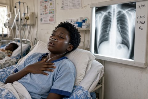
- Chest radiograph (erect, posteroanterior): consolidation, effusion, pneumothorax (visible lung edge), wedge infarct (Hampton hump), elevated hemidiaphragm, cardiomegaly, rib fracture. Lateral view or ultrasound for small effusion.
- Thoracic ultrasound at the bedside: absent lung sliding (pneumothorax), pleural fluid, B lines, pericardial effusion, consolidation. CT thorax for complex disease.
- Blood tests: full blood count, C reactive protein, procalcitonin, urea, creatinine, electrolytes, liver function tests, lactate, blood cultures, HIV test with consent, autoimmune screen (antinuclear antibody, anti double stranded DNA, rheumatoid factor, complement) and thrombophilia screen when indicated, lipase.
- Pleural effusion: diagnostic thoracentesis under ultrasound (protein, lactate dehydrogenase, glucose, pH, cell count and differential, Gram stain and culture, acid fast bacilli and GeneXpert, adenosine deaminase over 40 units/L supports tuberculosis, cytology).
- Light criteria for exudate: pleural protein to serum protein ratio over 0.5, lactate dehydrogenase ratio over 0.6, or pleural lactate dehydrogenase over two thirds of the upper limit of normal serum value.
- Pleural pH under 7.2 or glucose under 3.3 mmol/L indicates complicated effusion needing drainage.
- Sputum for culture and acid fast bacilli; pneumococcal and Legionella urinary antigens in severe pneumonia.
- Pericarditis: echocardiography, C reactive protein, troponin; tuberculous or malignant causes need pericardial fluid analysis.
- Rib radiograph is rarely helpful; CT or ultrasound for suspected occult rib fracture with complications.
Limits. D-dimer is nonspecific (infection, cancer, pregnancy, age). Chest radiographs are normal in about 25% of pulmonary emboli. Pleuritic pain with normal investigations is often viral or musculoskeletal.
Analgesia. Paracetamol 1 g orally every 6 hours (maximum 4 g; 3 g if frail or under 50 kg) with ibuprofen 400 mg every 8 hours with food (maximum 2.4 g) or naproxen 500 mg twice daily when no contraindication.
Avoid NSAIDs with suspected pulmonary embolism awaiting anticoagulation if bleeding risk is high, renal impairment, or peptic ulcer disease.
Severe pain: opioids such as morphine 2.5 to 5 mg intravenously titrated (avoid respiratory depression), intercostal nerve block or paravertebral block. Encourage deep breaths despite pain to prevent atelectasis.
Cause directed treatment.
- Pulmonary embolism: anticoagulation for at least 3 months, indefinite if unprovoked with low bleeding risk.
- Rivaroxaban 15 mg twice daily for 21 days then 20 mg daily, or apixaban 10 mg twice daily for 7 days then 5 mg twice daily.
- Enoxaparin 1 mg/kg twice daily or 1.5 mg/kg once daily; in renal impairment use unfractionated heparin with infusion adjusted to aPTT 1.5 to 2.5 times control.
- Pregnancy: enoxaparin 1 mg/kg twice daily.
- Thrombolysis (alteplase 100 mg over 2 hours) for massive embolism with hypotension.
- Pneumonia: amoxicillin 1 g orally three times daily for 5 days for mild disease.
- Moderate to severe: co-amoxiclav 1.2 g intravenously every 8 hours with clarithromycin 500 mg twice daily (or azithromycin 500 mg daily) for 5 to 7 days; severe: add oxygen, fluid, and escalate.
- Doxycycline 200 mg then 100 mg daily if penicillin allergic.
- Parapneumonic effusion and empyema: therapeutic drainage with chest drain for pH under 7.2, pus, or positive Gram stain; intrapleural alteplase 10 mg with DNase 5 mg twice daily for 3 days in selected patients; surgery for failure.
- Tuberculous pleurisy: isoniazid 5 mg/kg (maximum 300 mg), rifampicin 10 mg/kg (maximum 600 mg), pyrazinamide 25 mg/kg (maximum 2 g) and ethambutol 15 mg/kg daily for 2 months, then isoniazid and rifampicin for 4 months, with pyridoxine 10 mg daily; drain symptomatic effusion.
- Pneumothorax, tension: needle decompression then chest drain.
- Primary spontaneous with minimal symptoms and rim under 2 cm: observation.
- Larger or symptomatic: aspiration with a 16 to 18 G cannula (up to 2.5 litres) or small bore chest drain.
- Recurrent: surgical pleurodesis; advise no flying for 2 weeks after radiographic resolution and no diving permanently unless pleurodesis.
- Pericarditis: ibuprofen 600 mg every 8 hours for 1 to 2 weeks with taper and colchicine 500 micrograms twice daily (once daily if under 70 kg) for 3 months; gastroprotection; avoid anticoagulants if possible; prednisolone only for refractory disease.
- Tuberculous pericarditis needs anti tuberculous treatment plus prednisolone 60 mg daily tapered.
- Systemic lupus erythematosus and other autoimmune pleurisy: NSAIDs, prednisolone 0.5 mg/kg daily, hydroxychloroquine 200 to 400 mg daily, and rheumatology care. Familial Mediterranean fever: colchicine 1 to 2 mg daily.
- Malignant effusion: therapeutic drainage, indwelling pleural catheter or talc pleurodesis (4 to 5 g), oncology care.
- Chest wall causes: reassurance, heat, simple analgesia, rest; rib fracture analgesia and early mobilisation, with regional block for multiple fractures; costochondritis treated with NSAIDs and reassurance.
- Herpes zoster: valaciclovir 1 g three times daily for 7 days started within 72 hours, analgesia; for post herpetic neuralgia pregabalin 75 mg twice daily increasing to 150 mg twice daily (maximum 300 mg twice daily), or amitriptyline 10 to 25 mg at night.
- Pleurodynia: analgesia and heat; self limiting in 3 to 7 days.
- Sickle cell acute chest syndrome: oxygen, analgesia, incentive spirometry, antibiotics (ceftriaxone 2 g daily with azithromycin 500 mg daily), and transfusion for hypoxia; exchange transfusion in severe disease.
- Subdiaphragmatic disease: treat the source (antibiotics, drainage, surgery).
Monitoring. Respiratory rate, saturation, temperature and pain scores; repeat chest radiograph after 6 weeks for pneumonia and effusion in smokers or those over 50; repeat echocardiography for pericardial effusion; anticoagulation monitoring.
Referral and escalation. Respiratory medicine for effusion, empyema, recurrent pneumothorax or tuberculosis. Cardiology for pericarditis with effusion. Haematology for recurrent thromboembolism. Rheumatology for autoimmune causes. Cardiothoracic surgery for complicated empyema, persistent air leak or tamponade.
Admission.
- Haemodynamic instability, hypoxia.
- Pulmonary embolism with risk features (Pulmonary Embolism Severity Index class III or higher).
- Large or symptomatic pneumothorax.
- Pneumonia with CURB-65 score of 2 or more.
- Empyema.
- Pericarditis with effusion or high risk features (fever over 38 degrees Celsius, subacute course, large effusion, myopericarditis, immunosuppression).
- Severe pain, or social concerns.
Selected low risk pulmonary embolism may be treated as an outpatient.
Safety netting. Return for worsening breathlessness, haemoptysis, fever, collapse, leg swelling, rapidly increasing pain, or palpitations.
Special groups. Pregnancy: pulmonary embolism (V/Q scan or CT with shielding is acceptable), peripartum cardiomyopathy; enoxaparin only. Children: pneumonia, viral pleurisy, pneumothorax and sickle cell crisis. Older adults: atypical pneumonia, malignancy and embolism. Endemic regions: tuberculosis, amoebic liver abscess, hydatid disease and sickle cell disease.
