








Wound Healing
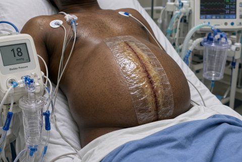
Wound healing is the coordinated restoration of tissue integrity after injury. It proceeds through overlapping phases, and failure at any stage produces a chronic wound, excessive scarring or dehiscence.
Phases
- Haemostasis (minutes to hours): vasoconstriction, platelet aggregation and fibrin clot formation. Platelet granules release PDGF, TGF-beta and fibronectin.
- Inflammation (days 1 to 4): neutrophils clear bacteria and debris in the first 48 hours, followed by macrophages (days 2 to 5), which orchestrate repair through growth factor release.
- Proliferation (days 4 to 21): fibroblasts deposit type III collagen, angiogenesis forms granulation tissue, keratinocytes migrate to re-epithelialise, and myofibroblasts drive contraction.
- Remodelling (3 weeks to 2 years): type III collagen is replaced by type I, cross-linking increases, and the scar reaches only about 70 to 80% of the original tensile strength.
Healing intentions
- Primary: edges apposed by sutures, staples or adhesive. Minimal scar.
- Secondary: left open to heal by granulation and contraction, used for contaminated wounds and abscess cavities.
- Tertiary (delayed primary): closure after 3 to 5 days once infection risk has passed.
Factors impairing healing
- Local: infection, ischaemia, tension, foreign body, necrotic tissue, haematoma, radiation, repeated trauma.
- Systemic: diabetes, malnutrition (protein, vitamin C, zinc), anaemia, smoking, advanced age, obesity, uraemia, jaundice, immunosuppression.
- Drugs: corticosteroids, chemotherapy, anticoagulants, NSAIDs, immunosuppressants, mTOR inhibitors.
Abnormal scarring
- Hypertrophic scars stay within the wound margin and regress over 1 to 2 years. Keloids extend beyond it, rarely regress and are commoner in darker skin. Contractures occur across joints.
- Chronic wounds fail to progress through healing within 4 to 6 weeks. Common types are venous, arterial, diabetic and pressure ulcers.
