









MRI Abdomen and Liver
MRI of the abdomen and liver is a multiparametric study of the liver, biliary tree, pancreas, spleen, kidneys, adrenals and retroperitoneum. It is the best non-invasive test for characterising focal liver lesions and for assessing diffuse liver disease.
The study uses a 1.5 or 3 tesla scanner with a phased array body coil. Slices are 4 to 6 mm thick. Most sequences are acquired in a breath hold of 15 to 20 seconds, and the whole study takes 30 to 45 minutes.
Core sequences are T1-weighted in phase and opposed phase (chemical shift imaging for fat and iron), T2-weighted with fat suppression, diffusion weighted imaging, and dynamic contrast enhanced T1 with fat suppression (arterial, portal venous, equilibrium and delayed phases). Hepatobiliary phase imaging uses gadoxetate (20 to 40 minutes after injection), and quantitative sequences measure liver fat, iron and stiffness.
Focal liver lesions are found in 20 to 30% of adults, and most are benign. MRI has a sensitivity of over 90% and a specificity of 90 to 95% for characterising lesions as benign or malignant. It detects liver metastases smaller than 1 cm better than CT, and diagnoses hepatocellular carcinoma in cirrhosis with a sensitivity of 80 to 90% through typical enhancement.
Chronic liver disease affects hundreds of millions of people (fatty liver in about 25% of adults, viral hepatitis, alcohol). MRI proton density fat fraction quantifies steatosis accurately, and iron overload is measured by T2* or R2 relaxometry.
Indications.
- Characterisation of indeterminate liver lesions found on ultrasound or CT.
- Surveillance and diagnosis of hepatocellular carcinoma in cirrhosis, and staging.
- Liver metastases detection before resection or ablation (colorectal cancer, neuroendocrine tumours).
- Cholangiocarcinoma and gallbladder cancer staging.
- Pancreatic lesions: solid masses, cystic neoplasms, neuroendocrine tumours, pancreatitis.
- Quantification of liver fat, iron and fibrosis (elastography).
- Budd-Chiari syndrome, portal vein thrombosis and vascular disease.
- Renal and adrenal masses that are indeterminate on CT or ultrasound (adrenal adenoma versus metastasis, phaeochromocytoma).
- Spleen lesions and lymph nodes.
- Abdominal wall and retroperitoneal masses.
- Inflammatory bowel disease, and appendicitis in pregnancy.
- Pre-operative planning for liver resection, live donor transplantation and ablation.
- Post-treatment response (ablation, embolisation, chemotherapy).
- Patients with contraindication to iodinated contrast or radiation.
Not for routine investigation of non-specific abdominal pain, or for patients who cannot hold their breath or lie still.
Contraindications and precautions.
- The usual MRI safety screening (pacemakers, clips, implants, metal fragments). Surgical clips in the abdomen cause local artefact.
- Gadolinium: avoid in severe renal impairment with linear agents. Macrocyclic agents are used at the lowest dose. Gadoxetate is also excreted via the bile and gives lower dynamic enhancement.
- Pregnancy: non-contrast MRI if needed.
- Claustrophobia, large body size exceeding the bore, and inability to hold the breath.
- Fasting for 4 to 6 hours reduces bowel motion and gallbladder contraction. Hyoscine or glucagon may be given.
- Oral contrast (negative agents or water) is optional for pancreatic and bowel studies.
Preparation and technique.
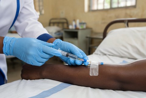
The patient fasts for 4 to 6 hours. A cannula is placed in the antecubital vein. The patient lies supine with the coil over the abdomen and learns the breath hold instructions.
T2 images are taken in axial and coronal planes, with fat suppression and respiratory triggering for free breathing sequences. Chemical shift (in- and opposed phase) images detect fat and iron. Diffusion weighted images are taken with b values of 50 and 800 or higher.
Dynamic contrast imaging uses a 3D gradient echo sequence with bolus timing, and arterial, portal venous and delayed phases. For gadoxetate, the transitional phase at 3 minutes and the hepatobiliary phase at 20 minutes are added.
MR elastography uses a passive driver on the chest wall to measure liver stiffness. Proton density fat fraction and T2* maps are acquired in a single breath hold each.
