











Abdominal Wall Hematoma
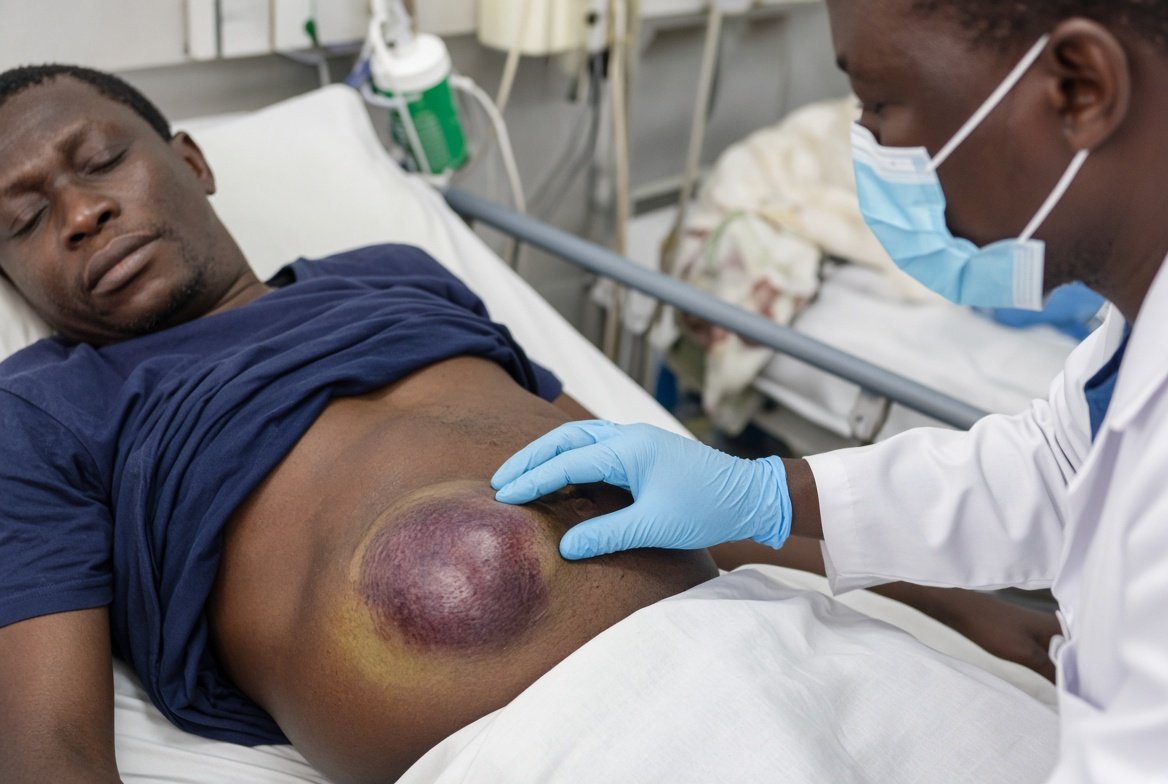
An abdominal wall hematoma is a collection of blood in the subcutaneous fat or in the muscle and fascial planes of the anterior and lateral abdominal wall, outside the peritoneal cavity.
It arises from injury to the superficial or deep epigastric vessels, the circumflex iliac vessels or small muscular perforators.
Causes
Blunt trauma (seat belt, handlebar, kick) and penetrating injury
Laparotomy and port wounds, paracentesis and drain insertion
Subcutaneous injections of enoxaparin, heparin or insulin
Anticoagulants, antiplatelet drugs, thrombocytopenia, hemophilia and acquired factor deficiency
Violent coughing or vomiting
Wound hematoma complicates 1 to 5% of abdominal operations, and risk rises with obesity, anticoagulation and hypertension.
Most small collections resorb over 2 to 6 weeks. The wall can hold 1 to 2 L before the patient looks unwell.
Complications
Expanding hematoma with hypovolemia
Infection and abscess
Skin necrosis and wound dehiscence
Chronic seroma
Abdominal compartment syndrome in massive bleeds
A seat belt sign (a linear bruise across the lower abdomen) marks deeper injury, with hollow viscus or mesenteric injury in roughly 20 to 30%, and a lumbar flexion-distraction fracture in some.
