








Emphysema Predominant COPD
COPD in which destruction of alveolar walls and loss of elastic recoil distal to the terminal bronchioles is the dominant lesion, causing hyperinflation, air trapping and impaired gas transfer, with relatively little chronic cough or sputum. Emphysema is found on CT in about 40 to 60% of people with COPD.
- Patterns: centrilobular (upper lobe, smoking), panlobular (lower lobe, alpha-1 antitrypsin deficiency), and paraseptal (subpleural, bulla formation). The "pink puffer" picture (thin, breathless, hyperinflated, with preserved oxygenation until late) is the typical severe form.
- Causes: tobacco smoking, cannabis smoking, biomass smoke, occupational dusts, intravenous drug use (talc), HIV infection, and alpha-1 antitrypsin deficiency (PiZZ phenotype, with onset before age 50 to 55, lower lobe disease and a family history).
- Prognosis is determined by FEV1, gas transfer (diffusing capacity), BMI, exercise capacity and exacerbation frequency. Hyperinflation and low BMI are strong predictors of death.
- Progressive exertional breathlessness, minimal cough, weight loss, muscle wasting, fatigue, reduced exercise tolerance.
- Signs: barrel chest, hyperinflation with reduced cardiac and liver dullness, quiet breath sounds, prolonged expiration, pursed lip breathing, use of accessory muscles, tripod posture, cachexia, hyperresonant percussion, low lying diaphragm. Cyanosis occurs late.
- Differential diagnosis: chronic bronchitis phenotype COPD, asthma, bronchiectasis, interstitial lung disease with emphysema (combined pulmonary fibrosis and emphysema), lymphangioleiomyomatosis, Langerhans cell histiocytosis, alpha-1 antitrypsin deficiency, bronchiolitis obliterans, heart failure, anaemia, deconditioning.
- Complications: bulla formation and pneumothorax, exacerbations, respiratory failure, pulmonary hypertension, cachexia and osteoporosis, lung cancer (risk raised 2 to 5 times), depression and anxiety.
- Spirometry: post-bronchodilator FEV1/FVC less than 0.70 with an irreversible pattern. Lung volumes: raised total lung capacity and residual volume (RV/TLC greater than 40 to 45%) show air trapping. Diffusing capacity for carbon monoxide is reduced (less than 80% predicted, often less than 50% in severe disease), a key difference from chronic bronchitis.
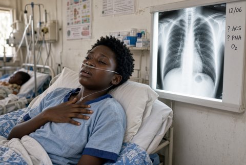
- High resolution CT: low attenuation areas (less than minus 950 Hounsfield units) distribution by lobe, bullae, and fissure completeness (needed for valve planning). Chest X-ray: hyperinflation, flat diaphragm, increased retrosternal space, narrow heart, bullae.
- Alpha-1 antitrypsin level in everyone with COPD (serum less than 11 micromol/L, or about 80 mg/dL, suggests deficiency) followed by phenotype or genotype testing; test first degree relatives.
- Walk test, cardiopulmonary exercise testing, arterial blood gas, echocardiography for pulmonary hypertension, BMI and body composition, assessment for lung cancer (annual low dose CT in eligible smokers).
- Smoking cessation (essential): varenicline 0.5 mg daily for 3 days, 0.5 mg twice daily for 4 days, then 1 mg twice daily for 12 weeks; nicotine patch 21 mg/24 h plus short acting nicotine replacement; bupropion 150 mg daily for 3 days then twice daily; behavioural support. Stop cannabis and other inhaled substances.
- Drug treatment:
- Long acting muscarinic antagonist and beta agonist combination: indacaterol/glycopyrronium 110/50 micrograms once daily, or umeclidinium/vilanterol 62.5/25 micrograms once daily, or tiotropium/olodaterol 5/5 micrograms 2 puffs once daily; salbutamol 100 to 200 micrograms as needed. Dynamic hyperinflation is reduced by long acting bronchodilators.
- Triple therapy (adds inhaled corticosteroid) for frequent exacerbations with eosinophils of 300 per microlitre or more, (fluticasone furoate/umeclidinium/vilanterol 100/62.5/25 micrograms daily).
- Alpha-1 antitrypsin augmentation therapy: IV alpha-1 proteinase inhibitor 60 mg/kg once weekly in non-smokers with deficiency (serum level less than 11 micromol/L), FEV1 35 to 65% predicted and established emphysema; it slows loss of lung density on CT, though evidence for clinical benefit is limited.
- Oral corticosteroids, theophylline and long term antibiotics have limited place; macrolide prophylaxis (azithromycin 250 mg daily) for frequent exacerbators who are ex smokers, after QT, hearing and mycobacterial checks.
- Pulmonary rehabilitation (twice weekly for 6 to 8 weeks, then maintenance): improves exercise capacity, breathlessness and quality of life; includes breathing retraining (pursed lip breathing, diaphragmatic breathing), inspiratory muscle training, upper limb training and education.
- Nutrition: high calorie, high protein diet (1.5 g/kg/day of protein), small frequent meals, oral supplements for BMI less than 21 kg/m2, and vitamin D with calcium when deficient.
- Oxygen: long term oxygen therapy for at least 15 hours daily when PaO2 is 7.3 kPa or less (or 8.0 kPa or less with cor pulmonale, haematocrit greater than 55% or nocturnal hypoxaemia); ambulatory oxygen for exertional desaturation to less than 88%; do not fly with SpO2 less than 92% without assessment.
- Home non-invasive ventilation for persistent hypercapnia (PaCO2 greater than 7 kPa) after an acidotic admission.
- Lung volume reduction in selected patients with severe hyperinflation and limited exercise capacity despite optimal medical therapy and rehabilitation:
- Surgical volume reduction (resection of the most diseased 20 to 30% of each lung): benefits patients with upper lobe predominant emphysema and low post-rehabilitation exercise capacity; avoid when FEV1 is less than 20% predicted with homogeneous emphysema or diffusing capacity less than 20% predicted (high mortality), or with pulmonary hypertension, hypercapnia (PaCO2 greater than 7.3 kPa) or age over 75. Operative mortality about 5%; 5-year survival improved in the right subgroup.
- Bronchoscopic valves (one way endobronchial valves): for FEV1 15 to 45% predicted, residual volume greater than 175% predicted, and a target lobe with an intact fissure (greater than 90% complete) shown by CT or a catheter based collateral ventilation measurement; improves FEV1 by about 100 to 200 mL, exercise capacity and quality of life, with pneumothorax in about 20 to 30% in the first days (admit for 3 to 5 days).
- Thermal vapour ablation and coils are options in specialist centres (evidence is limited).
- Lung transplantation: refer for FEV1 less than 25% predicted with rapid decline, repeated exacerbations with hypercapnia, pulmonary hypertension, or functional class IV breathlessness despite optimal care, in patients under 65 to 70 who have stopped smoking and have adequate nutrition and support; survival about 50% at 5 years.
- Vaccination: yearly influenza, pneumococcal conjugate, COVID-19 and respiratory syncytial virus (older adults).
- Pneumothorax: spontaneous pneumothorax in emphysema is serious (poor reserve): intercostal chest drain 14 to 20 French, with suction at 10 to 20 cmH2O if there is persistent air leak, and early surgical or bronchoscopic valve treatment for persistent leak beyond 3 to 5 days.
- Lung cancer screening with low dose CT for smokers aged 50 to 80 with 20 or more pack years where available.
- Palliation: low dose morphine (modified release 5 to 10 mg orally twice daily, or immediate release 2.5 mg every 4 hours as needed) for refractory breathlessness, a handheld fan, anxiolytics when needed, and advance care planning.
- Special groups:
- Alpha-1 antitrypsin deficiency: screen family, avoid smoking and hepatotoxins, monitor liver disease (cirrhosis in about 10 to 15% of PiZZ adults), and consider augmentation therapy in non-smokers.
- HIV: antiretroviral therapy, and look for opportunistic lung disease.
- Pregnancy: rare; inhaled bronchodilators and inhaled corticosteroid; assess for oxygen needs.
- Elderly: tailor rehabilitation, simplify inhalers, and address frailty and depression.
- Red flags: sudden breathlessness with chest pain (pneumothorax), haemoptysis (cancer, infection), weight loss greater than 5% in 6 months, hypercapnic drowsiness, right heart failure.
- Follow up: review every 3 to 6 months with spirometry yearly, weight and SpO2, exacerbation history; CT yearly for suspected nodules or when screening; specialist review for lung volume reduction or transplant assessment when criteria are met.
