Transmission. Airborne, via inhalation of small droplet nuclei, under 5 micrometres. An individual with active pulmonary or laryngeal tuberculosis produces them by coughing, speaking, or sneezing.
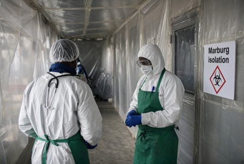
These droplet nuclei remain suspended in air for extended periods and can travel well beyond the immediate vicinity of the source. In the healthcare setting this requires airborne precautions: negative-pressure isolation room, and N95 or FFP respirator use by staff. These differ from the shorter-range droplet precautions used for many other respiratory pathogens.
Pathogenesis and granuloma formation. Inhaled organisms reach the alveoli and are phagocytosed by alveolar macrophages. The organism survives within them via the intracellular mechanism.
Over several weeks, a cell-mediated immune response develops, with T cell recognition of mycobacterial antigen and activation of macrophages. This leads to formation of a granuloma. It is an organised collection of activated macrophages, some fusing into multinucleated giant cells, surrounded by lymphocytes. It functions to contain, though not necessarily eliminate, the organism.
The centre of a mature tuberculous granuloma characteristically undergoes caseous necrosis, a soft, cheese-like necrotic centre. This gives the granuloma type its name, caseating granuloma. On tissue biopsy it is a distinctive histological feature strongly associated with tuberculosis. A small number of other specific granulomatous conditions less commonly show it.
Primary infection is the initial encounter between the organism and an immunologically naive host. It is typically in childhood in high-prevalence settings. It is most commonly clinically silent or associated with only mild, nonspecific symptoms.
The primary lung lesion, the Ghon focus, together with the associated hilar or mediastinal lymph node involvement, is termed the Ghon complex. This is a classic radiological and pathological finding at the site of primary infection. It may later calcify and remain visible on imaging indefinitely as a marker of prior exposure.
In the great majority of immunocompetent individuals, the developing cell-mediated immune response successfully contains primary infection. The organism enters a state of latency rather than progressing directly to active disease.
Latent infection is a state in which the organism persists within the granuloma in a dormant, non-replicating or minimally replicating form. Ongoing immune surveillance contains but does not eliminate it. It produces no clinical symptoms and is not infectious to others.
It is the outcome for the substantial majority of infected immunocompetent individuals. They carry a lifetime risk of later reactivation, estimated around 5 to 10 percent overall. The risk is concentrated disproportionately in the first two years after infection and further elevated by any subsequent immunosuppression.
Latent infection is detected by tuberculin skin testing (the Mantoux test) or interferon-gamma release assays.
- The Mantoux test measures a delayed-type hypersensitivity response to injected mycobacterial antigen. It can produce a false-positive result in a BCG-vaccinated individual.
- Interferon-gamma release assays are blood tests measuring T cell interferon-gamma release in response to specific M. tuberculosis antigens. Prior BCG vaccination does not confound them.
Reactivation occurs when the immune containment of latent infection fails. The most common cause is acquired immunosuppression. HIV infection is the single most important global risk factor, dramatically increasing both the risk and the speed of progression from latent to active disease.
Other associations are:
- Malnutrition, diabetes and chronic kidney disease.
- Certain immunosuppressive medications, notably tumour necrosis factor inhibitors used in various autoimmune conditions. This requires latent TB screening before starting such therapy.
- Advancing age.
Reactivation disease classically involves the lung apices (the Simon focus). The upper lobes have relatively higher regional oxygen tension, an environment favouring this obligate aerobe. This is distinct from the typically lower or mid-zone location of primary infection.
Pulmonary disease is the dominant clinical manifestation of active tuberculosis. It is a subacute-to-chronic illness with cough, often productive, and fever, night sweats, and weight loss. Haemoptysis is sometimes present in advanced or cavitary disease.
Cavitary disease is formation of an air-filled cavity within an area of lung necrosis. It is typically apical, reflecting reactivation disease specifically. It is associated with a particularly high mycobacterial burden and correspondingly high infectiousness. The necrotic, organism-rich cavity contents communicate directly with the airway.
Extrapulmonary disease occurs via haematogenous or lymphatic spread from the primary or reactivated pulmonary focus. It can involve essentially any organ system:
- Lymphadenitis is the most common extrapulmonary site, classically cervical, historically termed scrofula.
- Pleural disease: tuberculous pleural effusion.
- Skeletal disease, particularly vertebral tuberculosis, termed Pott disease. It causes vertebral body destruction and collapse, potentially with associated cord compression.
- Genitourinary disease.
- Tuberculous meningitis, typically subacute in onset. The characteristic CSF profile is lymphocytic pleocytosis, elevated protein, and low glucose.
- Miliary tuberculosis: widespread haematogenous dissemination producing numerous small, millet-seed-sized granulomatous lesions across multiple organs. It is a severe manifestation particularly associated with young children and immunocompromised hosts.
Diagnostic microbiology. Sputum smear microscopy (Ziehl-Neelsen or auramine-rhodamine stain) provides a rapid preliminary result. It is a key measure of infectiousness: a smear-positive patient is considered substantially more infectious than a smear-negative, culture-positive patient. Sensitivity is only moderate, and it cannot itself distinguish M. tuberculosis from nontuberculous mycobacteria.
Culture on specialised solid media such as Lowenstein-Jensen agar, or more rapidly in liquid culture systems, remains the diagnostic gold standard. It is critically required to obtain an isolate for drug susceptibility testing. It is slow, taking days to several weeks depending on the method.
Nucleic acid amplification testing (PCR-based), for example the widely used Xpert MTB/RIF assay, provides same-day results with high sensitivity. It can distinguish M. tuberculosis from nontuberculous mycobacteria. Xpert MTB/RIF also simultaneously screens for rifampicin resistance. It is an increasingly central tool in both diagnosis and initial resistance screening, particularly valuable in high-burden settings.
Drug resistance principles. Standard first-line treatment for drug-susceptible tuberculosis uses a combination of multiple agents, classically isoniazid, rifampicin, pyrazinamide, and ethambutol. The course is prolonged, typically 6 months total, with an initial intensive phase followed by a continuation phase.
Combination therapy is needed to prevent selection of resistant organisms. These would otherwise emerge readily under single-drug pressure, given the organism's large natural population size within an established infection and the inevitability of spontaneous resistance mutations arising within that population.
Multidrug-resistant tuberculosis (MDR-TB) is defined as resistance to at least both isoniazid and rifampicin, the two most potent first-line agents. It requires prolonged treatment with a more complex, less effective, and more toxic second-line drug regimen.
Extensively drug-resistant tuberculosis (XDR-TB) is a further, more severe level of resistance: MDR-TB plus resistance to key second-line drug classes. It is associated with markedly worse treatment outcomes and is a major and actively monitored global public health concern.
Poor treatment adherence (incomplete courses, erratic dosing) is the principal driver of acquired drug resistance. This underlies the widespread use of directly observed therapy (DOT) programmes in many tuberculosis control settings. Treatment administration is directly supervised to maximise adherence and minimise resistance emergence.