








Candida auris
Also known as: Candidozyma auris, C. auris, Multidrug Resistant Candida
Candida auris (Candidozyma auris in recent taxonomy) is a multidrug resistant yeast that colonises skin, persists on surfaces, and spreads between patients in healthcare settings, causing outbreaks of candidaemia and wound, ear, and urinary infection.
It was first isolated in 2009 from the ear canal of a patient in Japan (the Latin name refers to the ear) and emerged independently on several continents in the following years.
It is notifiable in many jurisdictions and is on the highest priority tier of fungal pathogens published by the World Health Organization.
Three properties make it unusual among Candida species: it colonises skin (axilla, groin, nares, and skin folds) for months, it survives for weeks on dry surfaces and equipment, and it often carries resistance to more than one antifungal class. Crude mortality of candidaemia is 30 to 60 percent in reported series, reflecting patients who are already critically ill.
| Clade | Region of first recognition | Features |
|---|---|---|
| I | South Asia (India, Pakistan), also Europe and the United States | High fluconazole resistance; invasive disease |
| II | East Asia (Japan, Korea) | Ear isolates; usually susceptible to fluconazole |
| III | Africa (South Africa) | Fluconazole resistant |
| IV | South America (Venezuela, Colombia) | Invasive disease; fluconazole resistant |
| V | Iran | Single report cluster; rare |
| VI | Singapore and Bangladesh | Recently described; limited data |
Clades differ by tens of thousands of single nucleotide variants, which shows near simultaneous emergence from separate origins rather than spread from a single source. Proposed contributors are climate warming (thermotolerance up to 42 degrees Celsius), antifungal use in agriculture and healthcare, and an environmental or marine niche that is not yet established.
Risk factors for infection are admission to intensive care, long term care with ventilation (ventilator units in particular), central venous catheters, broad spectrum antibacterial and antifungal exposure, diabetes, chronic kidney disease, recent surgery, and previous colonisation. Colonisation precedes most invasive infections, and transmission is by contaminated hands, shared equipment (thermometers, blood pressure cuffs, reusable probes), and the near patient environment.
The cells are oval, 2.5 to 5 micrometres, and occur singly or in pairs, with no true hyphae and no germ tubes, though pseudohyphae or aggregates form in some strains. Aggregating phenotypes have increased biofilm formation and skin adherence. The yeast grows at 37 to 42 degrees Celsius and in high salt (10 percent sodium chloride), which is used for selective enrichment in screening.
| Class | Resistance frequency | Mechanisms |
|---|---|---|
| Fluconazole | About 90 percent of isolates overall | ERG11 substitutions (Y132F, K143R, and VF125AL), TAC1B gain of function with efflux pump (CDR1) overexpression |
| Other azoles | Variable cross resistance | Same mechanisms; voriconazole MICs rise with fluconazole resistance |
| Amphotericin B | About 30 percent, with high variation by region | Sterol biosynthesis changes, reduced ergosterol |
| Echinocandins | About 2 to 7 percent, higher in some outbreaks | FKS1 hot spot mutations (S639F, S639P, and S639Y) |
| Flucytosine | Many isolates susceptible | Not routinely tested |
Pan resistant isolates, which are resistant to azoles, polyenes, and echinocandins, have been reported and are rare. Echinocandin resistance can arise during therapy, so repeat isolates are retested.
- Colonisation: the commonest state, found in axilla, groin, nares, rectum, and the respiratory tract. It persists for months, does not cause disease in most carriers, and is not treated.
- Candidaemia: fever and shock indistinguishable from other candidaemia, often with a retained central line, and frequent persistence of positive blood cultures.
- Other infections: urinary tract infection (catheter associated), wound and surgical site infection, bone and joint infection, otitis, burn wound infection, respiratory isolates (mostly colonisation), and intra-abdominal infection. Meningitis and endocarditis are rare.
Misidentification is the central laboratory problem. Older biochemical panels and automated identification databases misidentify Candida auris as Candida haemulonii, Candida duobushaemulonii, Candida famata, Candida sake, Candida lusitaniae, Rhodotorula glutinis, or Saccharomyces cerevisiae. Any yeast identified as one of these species from blood or a sterile site in a high risk setting is sent for confirmation.
| Method | Use |
|---|---|
| Matrix assisted laser desorption ionisation time of flight mass spectrometry with an updated database | Reliable identification in hours; the database must include Candida auris |
| Sequencing of the D1/D2 region of the large subunit ribosomal RNA gene or the internal transcribed spacer | Confirmatory identification and clade assignment |
| Chromogenic medium for Candida auris | Screening; colonies appear white to pale with a blue halo on one commercial medium; not specific |
| Polymerase chain reaction on skin swabs | Rapid colonisation screening where available |
| Enrichment broth with high salt, with dulcitol, and with antibacterials at 40 degrees Celsius | Selective screening culture from swabs |
| Susceptibility testing by broth microdilution | Required for every invasive isolate, with provisional breakpoints from public health agencies |
Screening specimens are composite swabs of the axilla and groin, with additional sites (nares, rectum, wounds, and urine from catheterised patients) in outbreaks. A patient is classed as clinical case or colonised depending on whether infection is present.
Provisional breakpoints indicating resistance, defined by public health agencies from limited data, are fluconazole 32 micrograms per mL or higher, amphotericin B 2 micrograms per mL or higher, caspofungin 2 or higher, and anidulafungin or micafungin 4 or higher. These are not clinical breakpoints and should be confirmed against current guidance.
Treatment is of clinical infection only. Colonisation is not treated with antifungals.
| Situation | Therapy |
|---|---|
| Candidaemia and invasive infection in adults | Echinocandin: caspofungin 70 mg intravenously on day 1 then 50 mg daily, or micafungin 100 mg daily (up to 150 mg daily has been used), or anidulafungin 200 mg on day 1 then 100 mg daily |
| Echinocandin resistance, non response, or persistent candidaemia | Liposomal amphotericin B 5 mg per kg once daily, alone or with an echinocandin |
| Pan resistant or refractory disease | Combination of liposomal amphotericin B, an echinocandin, and flucytosine 25 mg per kg every 6 hours (if the isolate is flucytosine susceptible), under specialist advice |
| Neonates and infants | Amphotericin B deoxycholate 1 mg per kg once daily, or an echinocandin where amphotericin cannot be used |
| Urinary tract infection | Remove the catheter; if treatment is needed, amphotericin B deoxycholate 0.3 to 0.6 mg per kg daily, because echinocandins and most azoles reach low urinary levels and fluconazole resistance is common |
| Central nervous system disease | Liposomal amphotericin B 5 mg per kg daily with flucytosine 25 mg per kg every 6 hours |
Central venous catheters are removed. The duration of therapy for candidaemia is 14 days from the first negative blood culture, with repeat blood cultures daily until clearance and extension for persistent culture positivity, metastatic infection, or neutropenia.
Rezafungin has activity in vitro and a long half life, and ibrexafungerp has in vitro activity, but neither has an established role in candidaemia from Candida auris outside trials or specialist programmes. Fosmanogepix remains investigational.
Olorofim and flucytosine monotherapy are not used.
Azole therapy fails in most isolates, and fluconazole is not used empirically in settings where Candida auris circulates.
Control of spread is the main intervention, and the following are standard elements.
- Detection and notification: laboratory capacity for accurate identification, immediate notification of infection control teams and public health authorities, and review of past isolates during outbreaks.
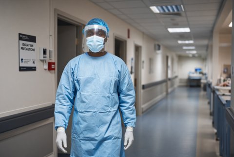
- Isolation: single room with its own bathroom or dedicated commode, contact precautions with gloves and gowns, and cohorting where single rooms are unavailable.
- Dedicated equipment: single patient or disinfected equipment, with particular care for reusable devices.
- Environmental cleaning: daily and terminal cleaning with a disinfectant active against Candida auris (chlorine based agents at 1000 parts per million or more of available chlorine, or products registered as effective against Candida auris), because quaternary ammonium agents alone are inadequate. Hydrogen peroxide vapour and ultraviolet light have been used as adjuncts.
- Hand hygiene: alcohol based hand rub and soap and water, with attention to contact points.
- Screening: admission screening of high risk patients (transfers from outbreak facilities or from regions with high prevalence, and those who have had overseas healthcare), and contact screening around new cases, repeated at intervals.
- Skin decolonisation: daily bathing with chlorhexidine gluconate 2 percent reduces skin burden, though recolonisation is common, and no regimen reliably eliminates carriage.
- Duration of precautions: carriage can persist for months, and many programmes continue precautions for the rest of an admission and apply them again at readmission, with release only after repeated negative screens under local policy.
- Antimicrobial stewardship: reduces selective pressure.
- Interfacility communication: transfer paperwork states the patient's colonisation status.
