










Tinea Corporis
Also known as: Ringworm
Tinea corporis is dermatophyte infection of glabrous skin of the trunk and limbs, excluding the groin, palms, soles and face. Tinea faciei is infection of the facial skin. Tinea imbricata is a distinctive chronic form caused by Trichophyton concentricum.
Organisms:
| Organism | Typical setting |
|---|---|
| Trichophyton rubrum | Commonest cause worldwide; adults, often with tinea pedis or onychomycosis as the reservoir |
| Trichophyton mentagrophytes and Trichophyton interdigitale | Inflammatory tinea; rodent, rabbit and guinea pig contact |
| Microsporum canis | Children, pet contact (kittens and puppies); ring lesions of the face and trunk |
| Trichophyton tonsurans | Contact sports (wrestling, tinea gladiatorum), household spread from scalp disease |
| Trichophyton verrucosum | Farmers and cattle workers |
| Epidermophyton floccosum | Occlusive clothing, groin to trunk spread |
| Nannizzia gypsea | Soil exposure, gardening |
| Trichophyton indotineae | Extensive, itchy, widespread disease, terbinafine resistant |
| Trichophyton concentricum | Tinea imbricata |
Risk factors: warm, humid climate, sweating and occlusive clothing, contact sports, close household or animal contact, obesity, diabetes mellitus, topical or systemic corticosteroids, and cellular immunodeficiency. Spread is by direct contact with an infected person, animal or soil, and by fomites such as towels, mats and clothing.
Hyphae spread outward in the stratum corneum at the rate of growth of the keratin layer, so the active edge advances while the centre clears as the host response and cell turnover eliminate the organism. This produces the annular morphology. Zoophilic species provoke neutrophilic and T cell responses, and the edge becomes vesicular or pustular, and may extend into hair follicles. Trichophyton rubrum provokes minimal inflammation, so lesions are scaly, chronic and often extensive.
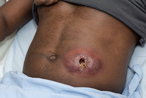
Topical corticosteroids suppress the inflammatory edge and reduce scale and itch, but permit fungal growth. The resulting lesion is called tinea incognito: ill defined borders, less scale, pustules or follicular papules, and spread over a wider area. Use of a combined corticosteroid and antifungal cream is a common cause.
Tinea corporis:
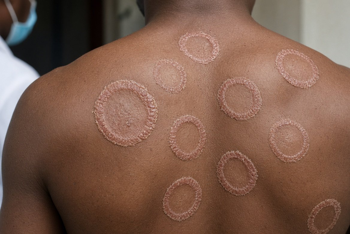
- One or more annular or arcuate plaques with a raised, erythematous, scaly edge, sometimes with vesicles, papules or pustules at the margin, and relative central clearing. The centre may stay scaly and pigmented.
- Itch is variable. Lesions enlarge slowly and may coalesce into polycyclic patches.
- Multiple lesions, large lesions or confluent plaques of the trunk, thighs and buttocks point to Trichophyton indotineae or immune compromise.
- Follicular involvement with papules and pustules indicates deeper infection of the hair follicle (Majocchi granuloma), which needs oral treatment.
- Children with zoophilic disease have inflamed, sometimes multiple lesions on exposed skin.
- Bullous tinea corporis is a rare presentation.
- Tinea gladiatorum is tinea corporis in contact sport athletes, usually on the head, neck and arms, caused by Trichophyton tonsurans, and spreads in teams.
Tinea faciei:
- Erythematous, scaly, often poorly demarcated patches of the cheeks, forehead, perioral and periorbital skin, and chin. Annular morphology is often absent.
- Itch, burning and photosensitivity make the lesions mimic lupus or rosacea. Sun exposure exacerbates redness.
- Facial involvement in men with beard hair is tinea barbae. Eyebrow, eyelash and scalp margin disease are related.
- Delay is common because of misdiagnosis as eczema, seborrhoeic dermatitis or rosacea and treatment with topical corticosteroids.
Tinea imbricata (Tokelau):
- Chronic infection by Trichophyton concentricum, endemic in the Pacific Islands (Tokelau, Samoa), Southeast Asia, the Indian subcontinent, Papua New Guinea, Central America and parts of South America.
- Concentric rings of overlapping, thin, lamellar scale, attached at their peripheral edge, produce a distinctive pattern of whorls and ripples. Lesions are extensive, covering the trunk and limbs, and may be widespread.
- Itch is intense, inflammation is minimal, hair is rarely involved, and nails are sometimes involved. Onset is in childhood and the course is chronic and relapsing for decades.
- Familial clustering is described, and an autosomal recessive susceptibility has been proposed. Infection reflects a poor delayed type hypersensitivity response.
- Wood lamp is negative. Potassium hydroxide shows hyphae. Culture of Trichophyton concentricum is slow and the colony is glabrous and heaped, and growth is easier on enriched media.
| Condition | Discriminating features |
|---|---|
| Nummular eczema | Coin shaped, oozing, crusted plaques; diffuse itch; no active edge with central clearing; KOH negative |
| Psoriasis | Thick silvery scale, extensor surfaces, nail pitting |
| Pityriasis rosea | Herald patch, oval lesions along skin cleavage lines with collarette scale |
| Granuloma annulare | Smooth, non scaly annular ring of papules |
| Erythema annulare centrifugum | Trailing scale behind an advancing edge; no hyphae |
| Erythema migrans | Expanding erythema, tick exposure; no scale |
| Subacute cutaneous lupus erythematosus | Photodistribution, annular or papulosquamous; serology |
| Impetigo | Honey coloured crust |
| Seborrhoeic dermatitis, rosacea, contact dermatitis, perioral dermatitis | Central face; no active scaly edge |
| Pityriasis versicolor | Fine scale in sebum rich skin; spaghetti and meatballs pattern |
| Ichthyosis and mycosis fungoides | Generalised scale or chronic patches unresponsive to antifungals |
- Specimen: scale from the active edge, taken with a blade after cleaning the skin with alcohol. A vesicle roof or pustule contents may be examined.
- Potassium hydroxide preparation: septate, branching hyphae of uniform width, sometimes with arthroconidia. Calcofluor white improves sensitivity.
- Culture: indicated for extensive or recurrent disease, tinea faciei, failure to respond, when oral therapy is planned, and for species identification. It takes 1 to 4 weeks.
- Molecular testing and sequencing: needed where resistance is suspected.
- Histopathology: hyphae in the stratum corneum on periodic acid Schiff, with sandwich sign.
- Pitfalls: recent topical antifungal or corticosteroid reduces the yield; scale from the centre of a lesion often contains no organisms.
Topical therapy
Topical treatment suffices for localised disease. The drug is applied to the lesion and to 2 cm of surrounding skin, and continued for 1 to 2 weeks after the lesion has cleared to reduce relapse.
| Agent | Regimen |
|---|---|
| Terbinafine 1 percent cream | Once daily for 1 week, or twice daily for 1 to 2 weeks |
| Naftifine 1 percent cream | Once daily for 2 weeks |
| Butenafine 1 percent cream | Once daily for 2 weeks |
| Luliconazole 1 percent cream | Once daily for 1 week |
| Clotrimazole 1 percent cream | Twice daily for 2 to 4 weeks |
| Miconazole 2 percent cream | Twice daily for 2 to 4 weeks |
| Econazole 1 percent cream | Once daily for 2 to 4 weeks |
| Ketoconazole 2 percent cream | Once daily for 2 to 4 weeks |
| Sertaconazole 2 percent cream | Twice daily for 4 weeks |
| Ciclopirox 0.77 percent cream | Twice daily for 4 weeks |
Allylamines and butenafine achieve mycological cure in a shorter time than azoles. Combined corticosteroid and antifungal creams are avoided, because they produce tinea incognito and worsen outcomes. Where itching is intense, an oral antihistamine is used instead.
Oral therapy
Oral therapy is indicated for extensive disease, multiple sites, folliculitic involvement, failure of topical therapy, chronic Trichophyton rubrum infection, tinea of the face in some cases, and immunosuppressed hosts.
| Agent | Adult dose | Duration |
|---|---|---|
| Terbinafine | 250 mg once daily | 1 to 2 weeks for skin disease; longer if slow response |
| Itraconazole | 100 mg once daily, or 200 mg once daily | 2 weeks at 100 mg daily, or 1 week at 200 mg daily |
| Fluconazole | 150 to 200 mg once weekly | 2 to 4 weeks |
| Griseofulvin (microsize) | 500 mg daily, in one or two doses | 2 to 4 weeks |
Children: terbinafine by weight (under 20 kg: 62.5 mg daily; 20 to 40 kg: 125 mg daily; over 40 kg: 250 mg daily) for 1 to 2 weeks, or griseofulvin 10 to 20 mg per kg daily for 2 to 4 weeks.
Pregnancy and breastfeeding: topical clotrimazole, miconazole or terbinafine. Oral terbinafine is avoided unless benefit outweighs risk, griseofulvin is contraindicated, and fluconazole is avoided in the first trimester.
Terbinafine resistant disease (Trichophyton indotineae)
- Terbinafine at standard dose and duration commonly fails.
- Itraconazole 200 mg daily for at least 4 weeks, usually 6 to 8 weeks or until clinical and mycological clearance, is the usual treatment. Some clinicians give 100 mg twice daily. Drug level monitoring can confirm adequate exposure. Relapse is frequent.
- Topical azole or ciclopirox is used as an adjunct. Susceptibility testing and SQLE sequencing guide treatment where available.
- Household and close contacts are examined, and shared fomites are decontaminated.
Tinea imbricata
Treatment is difficult and relapse is frequent. Oral terbinafine 250 mg daily for 4 weeks, itraconazole 100 mg daily for 4 weeks, or griseofulvin 500 mg to 1 g daily for several weeks achieve clearance in many patients, with limited trial evidence. Topical keratolytics (salicylic acid) and antifungals are added, and household members are examined and treated.
Prevention and control
- Treat tinea pedis, onychomycosis and tinea cruris, which reseed trunk disease.
- Examine household members and pets. Infected animals need veterinary treatment.
- Dry skin thoroughly, avoid occlusive clothing, and avoid sharing towels and razors.
- Contact sport teams: skin checks before practice, shower after practice, and wash mats and equipment. Prophylactic fluconazole 200 mg once weekly during the season reduces tinea gladiatorum in outbreak settings.
