








Extrapulmonary Aspergillosis
Also known as: Aspergillus Sinusitis, Cerebral Aspergillosis, Otomycosis, Cutaneous Aspergillosis
Extrapulmonary aspergillosis is infection of any site outside the lung parenchyma by Aspergillus. It has a broad spectrum from non invasive colonisation to rapidly fatal disease. The host, the site, and the species determine treatment.
Routes of infection:
- Haematogenous spread from the lung.
- Extension from adjacent infected tissue (sinuses to brain and orbit).
- Direct inoculation (surgery, trauma, catheters, contaminated dressings, and burns).
- Environmental contamination of the ear and eye.
Aspergillus flavus is the commonest cause in the sinuses, eye, skin, and central nervous system in hot climates.
| Site | Typical host or setting | Key features | Core treatment |
|---|---|---|---|
| Sinuses (invasive) | Neutropenia, transplant, diabetes, corticosteroids | Fever, facial pain, black eschar, orbital and cranial extension | Surgical debridement and voriconazole or liposomal amphotericin B |
| Sinuses (chronic granulomatous invasive) | Immunocompetent, Sudan, India, Pakistan, Saudi Arabia (Aspergillus flavus) | Proptosis, a slowly growing orbital or paranasal mass | Surgery and itraconazole for months |
| Sinus fungus ball | Immunocompetent adults | Maxillary sinus, unilateral | Endoscopic removal |
| Central nervous system | Haematogenous or sinus extension; neurosurgery; neutropenia, transplant | Abscess, stroke, mycotic aneurysm, meningitis | Voriconazole with neurosurgical drainage |
| Skin and soft tissue | Burns, neonates, catheter sites, dressings, neutropenia | Necrotic papules and eschars | Debridement and voriconazole |
| Eye | Keratitis (trauma, contact lens, corticosteroids); endophthalmitis (surgery, endogenous) | Corneal ulcer with feathery margins; vitritis | Natamycin and voriconazole; intravitreal injection and vitrectomy |
| Ear (otomycosis) | Warm humid climates, ear canal manipulation, chronic otitis | Itching, otorrhoea, fungal debris | Aural toilet and topical antifungal |
| Heart | Prosthetic valves, cardiac surgery, intravenous drug use | Culture negative endocarditis with large vegetations and emboli | Surgery with lifelong azole suppression |
| Bone and joint | Postoperative, trauma, chronic granulomatous disease | Vertebral osteomyelitis, sternal and prosthetic infection | Debridement and prolonged voriconazole |
| Gastrointestinal, hepatic, and renal | Neutropenia, transplant | Ulceration, abscess, perforation | Surgery and systemic antifungal therapy |
| Peritoneum | Peritoneal dialysis | Cloudy effluent | Catheter removal and antifungal therapy |
| Disseminated disease | Neutropenia, transplant | Multiorgan involvement and sepsis | Voriconazole or liposomal amphotericin B |
Acute invasive fungal rhinosinusitis
Occurs in neutropenia, haematological malignancy, transplant, and uncontrolled diabetes. Aspergillus flavus and Aspergillus fumigatus are the usual causes, and Mucorales must be excluded.
Presentation is fever, facial or periorbital pain, nasal congestion with black or necrotic mucosa, epistaxis, and visual loss. Spread to the orbit, cavernous sinus, and brain is rapid.
The diagnosis needs nasal endoscopy with biopsy of necrotic tissue for histology (acute angle branching septate hyphae) and culture, and imaging with computed tomography or magnetic resonance imaging.
Treatment is urgent surgical debridement, with voriconazole (6 mg per kg every 12 hours for 2 doses, then 4 mg per kg every 12 hours), or liposomal amphotericin B 5 mg per kg daily when mucormycosis is possible. Correct neutropenia and hyperglycaemia and reduce immunosuppression. Treatment continues for at least 6 to 12 weeks and until neutropenia and clinical findings resolve.
Chronic invasive and chronic granulomatous sinusitis
Aspergillus flavus chronic granulomatous invasive disease affects immunocompetent people in Sudan, India, Pakistan, and the Middle East. It causes a slowly growing mass in the maxillary, ethmoid, or sphenoid sinus, proptosis, and visual loss, resembling a tumour. Histology shows granulomas with giant cells and fungal hyphae.
Chronic invasive disease affects mildly immunosuppressed people (diabetes, corticosteroids). Treatment is surgery plus itraconazole 200 mg twice daily (or voriconazole) for at least 6 to 12 months, with level monitoring.
Fungus ball
A non invasive mass of hyphae and debris in a sinus (maxillary most often) of an immunocompetent adult, with unilateral symptoms (obstruction, discharge, facial pain). Computed tomography shows a hyperdense opacity with calcification. Treatment is endoscopic removal and ventilation of the sinus, and antifungals are not needed.
Cerebral aspergillosis arises from haematogenous seeding (the commonest route, in neutropenia and transplant), from direct extension from the sinuses, orbit, or middle ear, and after neurosurgery or lumbar puncture contamination.
Lesions are single or multiple abscesses, haemorrhagic infarcts (angioinvasion), mycotic aneurysms, meningitis, and granulomas. Presentation includes headache, fever, seizures, focal deficits, and altered consciousness. It is often silent early in neutropenia. Mortality is 80 to 90 percent in disseminated disease and lower after surgical treatment.
- Diagnosis: magnetic resonance imaging (ring enhancing lesions, restricted diffusion, haemorrhage, and infarcts), cerebrospinal fluid galactomannan, culture and polymerase chain reaction, serum galactomannan, and biopsy or aspiration of an accessible lesion. Cerebrospinal fluid galactomannan has a sensitivity of about 80 percent, and a positive result supports the diagnosis.
- Treatment: voriconazole 6 mg per kg every 12 hours for 2 doses then 4 mg per kg every 12 hours. It penetrates the brain well (trough 2 to 5.5 micrograms per mL targeted).
- Liposomal amphotericin B 5 mg per kg daily is an alternative or for resistant isolates.
- Neurosurgical drainage or resection of abscesses, and control of the source (sinus surgery).
- Combination therapy with an echinocandin or flucytosine is used in refractory cases, with limited evidence.
- The duration is at least 6 to 12 weeks and until radiological resolution, then secondary prophylaxis during further immunosuppression.
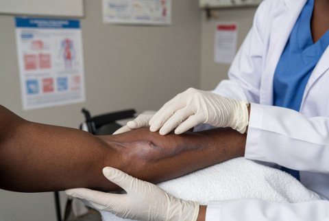
Primary cutaneous disease follows skin breach in immunocompromised and critically ill patients. Sources include intravenous catheter sites, arterial lines, adhesive tapes, occlusive dressings, burns, surgical wounds, and neonates (especially premature infants).
It presents as necrotic papules, plaques with a black eschar, or ulcers, with local invasion that may spread. Secondary disease is haematogenous from the lung.
Diagnosis is by biopsy with histology and culture, and a search for dissemination.
Treatment is surgical debridement, removal of the focus (catheter and dressing), and voriconazole or liposomal amphotericin B for at least 6 weeks (more in deep or disseminated disease), with correction of the host defect. Superficial primary cutaneous aspergillosis in immunocompetent patients may respond to debridement and topical azole.
| Syndrome | Features | Treatment |
|---|---|---|
| Fungal keratitis | Corneal ulcer with feathery edges, satellite lesions, hypopyon, after trauma (vegetable matter), contact lens wear, or topical corticosteroid; Fusarium and Aspergillus are the commonest filamentous causes | Natamycin 5 percent eye drops, hourly while awake for the first 1 to 2 weeks (then tapered); voriconazole 1 percent drops as an alternative or addition (natamycin was more effective than voriconazole in a randomised trial in Fusarium keratitis); intrastromal voriconazole 50 micrograms in 0.1 mL for deep disease; oral voriconazole for severe or deep disease; keratoplasty for perforation or failure |
| Endogenous endophthalmitis | Haematogenous spread in neutropenia or intravenous drug use, with vitritis and chorioretinal lesions | Systemic voriconazole, intravitreal voriconazole 100 micrograms in 0.1 mL or amphotericin B deoxycholate 5 to 10 micrograms in 0.1 mL, and early vitrectomy |
| Postoperative endophthalmitis | After cataract or other eye surgery | Intravitreal antifungal therapy, vitrectomy, and systemic voriconazole |
| Orbital aspergillosis | Spread from the sinuses; proptosis and visual loss | Surgical debridement and systemic antifungal therapy |
Otomycosis is superficial fungal infection of the external ear canal in warm and humid climates. It is caused by Aspergillus niger (the commonest, black spores), Aspergillus flavus, Aspergillus fumigatus, and Candida species.
Predisposing factors are ear canal manipulation, swimming, hearing aids, chronic otitis media, topical antibacterials and corticosteroids, and diabetes.
Presentation is itching, fullness, hearing loss, discharge, and pain, with white, grey, or black fungal debris resembling wet newspaper or fuzz.
Treatment is aural toilet (suction or microscopy guided cleaning), keeping the ear dry, and topical clotrimazole 1 percent solution or cream three times daily for 2 to 3 weeks. Alternatives are miconazole, econazole, ketoconazole drops, and acetic acid or boric acid solutions.
Invasive (malignant) external otitis from Aspergillus in diabetic and immunocompromised patients needs systemic voriconazole for months, with surgical debridement.
Aspergillus endocarditis (prosthetic valve and native valve), myocarditis, and pericarditis occur after cardiac surgery, in intravenous drug users, in neutropenia, and with disseminated disease. Endocarditis is typically culture negative (blood cultures positive in under 10 percent), with large vegetations, major arterial emboli, and mortality above 50 percent. Galactomannan and polymerase chain reaction help, and histology of emboli or valve tissue is diagnostic.
- Treatment: valve replacement and debridement, combined with voriconazole (6 mg per kg every 12 hours for 2 doses, then 4 mg per kg every 12 hours) or liposomal amphotericin B 3 to 5 mg per kg daily, for at least 6 weeks after surgery.
- Many patients then need lifelong oral azole suppression (voriconazole 200 mg twice daily, or itraconazole).
- Pericarditis and myocarditis: surgical drainage and prolonged antifungal therapy.
- Osteomyelitis and arthritis: vertebral osteomyelitis (often after surgery or in chronic granulomatous disease with Aspergillus nidulans), sternal osteomyelitis after cardiac surgery, rib and skull infection by extension, and prosthetic joint infection.
- Treatment is surgical debridement, removal of hardware where possible, and voriconazole for 6 to 12 months or longer, with duration guided by imaging and inflammatory markers.
- Gastrointestinal and hepatic aspergillosis: rare, with ulcers, perforation, and abscess in neutropenia and transplant; surgery and voriconazole.
- Renal and genitourinary aspergillosis: abscesses, papillary necrosis, and ureteric fungus balls with obstruction. Treat with drainage, relief of obstruction, and voriconazole, or amphotericin B. Urinary concentrations of voriconazole are low, but tissue levels are effective.
- Peritoneal dialysis peritonitis: catheter removal and voriconazole or amphotericin B.
- Tracheobronchitis and bronchial anastomosis infection: airway disease needs bronchoscopic debridement and voriconazole, and extension to the mediastinum needs surgical debridement.
- Mediastinitis and sternal wound infection: debridement and voriconazole.
- Nail and skin infection: Aspergillus species in onychomycosis (Aspergillus versicolor, Aspergillus terreus) need confirmation by repeated culture, and treatment with terbinafine or itraconazole.
- Disseminated aspergillosis: multiorgan involvement in neutropenia, haematogenous spread from a pulmonary primary; voriconazole or liposomal amphotericin B, with high mortality.
| Test | Role |
|---|---|
| Tissue biopsy with Grocott methenamine silver and periodic acid Schiff stain | Defines invasion; hyphae with acute angle branching and septa; negative stains with positive culture suggest contamination |
| Culture of tissue and fluid | Species identification, and susceptibility testing; sinus swabs are poor specimens compared with tissue |
| Galactomannan | Serum; cerebrospinal fluid, and other fluids (bronchoalveolar lavage); sensitivity high in neutropenic patients |
| Polymerase chain reaction | Tissue, cerebrospinal fluid, and blood; adds sensitivity |
| Beta D glucan | Supportive |
| Imaging | Computed tomography and magnetic resonance imaging define extension (orbit, brain, bone, and vessels) |
| Specialised tests | Fundoscopy, echocardiography, and Doppler ultrasonography as indicated |
Mucormycosis, fusariosis, scedosporiosis, and other mould infections look similar on histology and culture and need exclusion, because treatment differs.
| Principle | Detail |
|---|---|
| Surgery | Debridement is often needed for sinus, central nervous system, bone, cutaneous, and cardiac disease; antifungal drugs alone fail in necrotic tissue |
| First line drug | Voriconazole (or isavuconazole) for most sites; liposomal amphotericin B where mucormycosis cannot be excluded, azole resistance is possible, or an azole is contraindicated |
| Duration | Many weeks to months; chronic and device related disease needs the longest courses |
| Host recovery | Correcting neutropenia, hyperglycaemia, and corticosteroid exposure is part of treatment |
| Monitoring | Drug levels, liver tests, interactions, and repeated imaging |
| Secondary prophylaxis | Azole cover during further chemotherapy or transplantation |
