








Coccidioidomycosis
Also known as: Valley Fever, San Joaquin Valley Fever, Coccidioides Infection
Coccidioidomycosis (valley fever) is a systemic infection caused by the dimorphic fungi Coccidioides immitis and Coccidioides posadasii, acquired by inhaling airborne arthroconidia from desert soil. About 60 percent of infections are asymptomatic.
The rest present as a self limited community acquired pneumonia like illness, with a minority developing chronic pulmonary disease or extrapulmonary dissemination (1 to 5 percent), which can involve skin, bone, joints, and the meninges.
It is one of the most infectious fungi known, and cultures are handled at biosafety level 3.
| Feature | Description |
|---|---|
| Species | Coccidioides immitis (California, mainly the San Joaquin Valley, and Washington State) and Coccidioides posadasii (Arizona, New Mexico, Texas, Mexico, Central and South America) |
| Saprophytic phase | Hyaline septate hyphae in soil that fragment into barrel shaped arthroconidia (2 to 4 by 3 to 6 micrometres) separated by empty disjunctor cells; arthroconidia become airborne when soil is disturbed |
| Parasitic phase | Inhaled arthroconidia enlarge to spherules (20 to 80 micrometres, thick walled), which undergo internal cleavage and release endospores (2 to 5 micrometres); each endospore forms a new spherule |
| Culture | Fast growing white to grey cottony mould in 3 to 7 days on Sabouraud dextrose agar; arthroconidia are an extreme laboratory hazard, and the laboratory is told when coccidioidomycosis is suspected |
| Identification | DNA probe, sequencing, and mass spectrometry; the exoantigen test is replaced by molecular identification |
| Aspect | Details |
|---|---|
| Geography | The southwestern United States (Arizona and California account for most cases, with Nevada, Utah, New Mexico, and Texas), northern Mexico, and arid areas of Central and South America (Guatemala, Honduras, Venezuela, Colombia, Brazil, Paraguay, Bolivia, and Argentina); the endemic range is expanding northwards with climate change |
| Incidence | Over 20 000 cases are reported yearly in the United States, with under diagnosis estimated at 5 to 10 fold; incidence is highest in Arizona |
| Ecology | Alkaline desert soil with sparse vegetation; cases peak after dry spells followed by rain, with dust storms and earth moving |
| Exposure | Construction and agricultural work, military training, archaeological digs, wildfire and dust, and outdoor recreation; travellers can present after leaving the region |
| Dissemination risk | Filipino, African, Native American, and Hispanic ancestry; pregnancy (third trimester and postpartum); HIV with CD4 below 250; solid organ transplant; tumour necrosis factor inhibitors and corticosteroids; diabetes; male sex; age above 55 or in infancy |
| Transmission | Inhalation; person to person spread does not occur except in transplant (donor derived) and rare wound contamination |
Arthroconidia reach the alveoli and convert to spherules, which release endospores and induce a neutrophilic then granulomatous response. Cell mediated immunity (T helper 1 and T helper 17 responses with interferon gamma and interleukin 17) contains infection in most people and confers lasting immunity, with a positive skin test (spherulin). Defects in interferon gamma and interleukin 12 signalling, and in the STAT3, IL12RB1, and CARD9 pathways, predispose to dissemination. Reactivation occurs under later immunosuppression.
| Form | Features |
|---|---|
| Asymptomatic | About 60 percent |
| Acute pulmonary infection | After an incubation of 1 to 3 weeks: fatigue, cough, fever, pleuritic chest pain, night sweats, headache, and weight loss for weeks, with lobar or segmental consolidation, hilar adenopathy, and pleural effusion; peripheral eosinophilia in some; a leading cause of community acquired pneumonia in endemic regions (15 to 30 percent) |
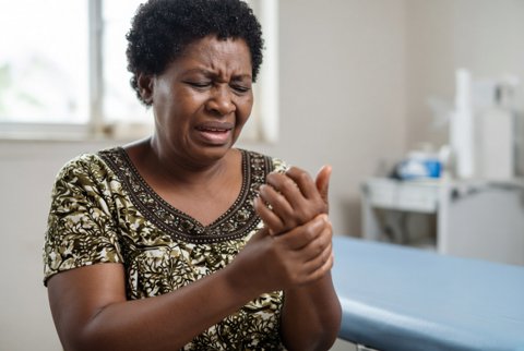
| Immune phenomena | Erythema nodosum (mostly women), erythema multiforme, arthralgia and arthritis ("desert rheumatism"), and a diffuse rash, which indicate good immunity |
| Pulmonary nodule | A solitary coin lesion, often found incidentally, and may mimic cancer |
| Pulmonary cavity | A thin walled cavity, which resolves in most, though it causes haemoptysis, infection, or rupture with pyopneumothorax |
| Chronic fibrocavitary disease | Chronic cough, weight loss, and haemoptysis, with upper lobe cavities that mimic tuberculosis |
| Diffuse pneumonia | Severe infection with acute respiratory distress syndrome, in immunosuppressed hosts and after heavy exposure |
| Skin (disseminated) | Verrucous plaques, nodules, ulcers, abscesses, and papules, most often on the face |
| Bone and joint | Vertebral, skull, and long bone osteomyelitis with cold abscesses, and arthritis of the knee and ankle |
| Meningitis | A subacute or chronic basal meningitis with headache, confusion, and cranial nerve palsies; hydrocephalus, vasculitis, and stroke; fatal without treatment |
| Others | Lymph nodes, prostate, liver, spleen, adrenal, genitourinary, ocular, and pericardial disease |
| Pregnancy | Severe disease in the third trimester and postpartum, with neonatal infection rare |
| Condition | Points that separate it |
|---|---|
| Bacterial and viral community acquired pneumonia | Failure to respond to antibacterials, history of travel and dust, and positive serology |
| Tuberculosis | Acid fast bacilli |
| Lung cancer and lymphoma | Histology |
| Histoplasmosis and blastomycosis | Morphology of the organism; antigen cross reaction |
| Sarcoidosis, granulomatosis with polyangiitis | Histology and autoantibodies |
| Cryptococcal meningitis, tuberculous meningitis, and carcinomatous meningitis | Cerebrospinal fluid antibody and culture |
| Test | Details |
|---|---|
| Serology | Enzyme immunoassay for IgM and IgG (a screening test with false positive IgM), then immunodiffusion for IgM (tube precipitin) and IgG (complement fixing antibody) and quantitative complement fixation to confirm; titres of 1 in 16 or higher indicate dissemination risk, and a rising titre is concerning; antibodies are absent in early disease and may be absent in the immunocompromised |
| Microscopy and histology | Calcofluor white, potassium hydroxide, Papanicolaou, or methenamine silver show spherules with endospores in sputum, bronchoalveolar lavage, pus, and tissue; spherules are pathognomonic |
| Culture | Positive in 20 to 50 percent of pulmonary cases; the laboratory is informed |
| Antigen | An enzyme immunoassay for coccidioidal antigen in urine, serum, or cerebrospinal fluid has about 70 percent sensitivity in disseminated disease, with cross reaction with other endemic fungi |
| Molecular tests | Polymerase chain reaction on respiratory and tissue specimens |
| Cerebrospinal fluid | Lymphocytic (and sometimes eosinophilic) pleocytosis, low glucose, high protein, and complement fixing antibody (diagnostic), with culture seldom positive |
| Imaging | Chest radiograph or computed tomography; magnetic resonance imaging of the spine and brain |
| Skin test | Spherulin test indicates past exposure and does not diagnose acute disease |
| Form | Regimen |
|---|---|
| Uncomplicated acute pulmonary disease | Most patients recover without antifungal therapy; treatment is considered in those with severe symptoms, high risk (diabetes, immunosuppression, pregnancy outside the first trimester, older age), or symptoms lasting longer than 2 months: fluconazole 400 mg once daily (400 to 800 mg), or itraconazole 200 mg twice daily, for 3 to 6 months |
| Severe, diffuse, or rapidly progressive pulmonary disease | Liposomal amphotericin B 3 to 5 mg per kg once daily (deoxycholate 0.5 to 1 mg per kg once daily) until improvement, then fluconazole 400 mg once daily or itraconazole 200 mg twice daily for at least 1 year |
| Chronic fibrocavitary disease | Fluconazole 400 mg once daily or itraconazole 200 mg twice daily for at least 12 months; surgery for haemoptysis, persistent cavities, and rupture |
| Extrapulmonary non meningeal disease | Fluconazole 400 to 800 mg once daily or itraconazole 200 mg twice daily for at least 1 year (bone disease often longer, with itraconazole preferred), with surgical debridement; amphotericin B for severe or vertebral disease |
| Meningitis | Fluconazole 800 to 1200 mg once daily (the usual 800 mg), for life, because relapse is frequent after stopping; itraconazole and voriconazole are alternatives; intrathecal or intraventricular amphotericin B deoxycholate (0.01 to 0.5 mg, with a reservoir) for failure or relapse; a ventriculoperitoneal shunt for hydrocephalus; corticosteroids may help vasculitis |
| HIV | Fluconazole 400 mg once daily (or amphotericin B for severe disease), with antiretroviral therapy, and lifelong suppression with fluconazole 200 to 400 mg daily until CD4 count above 250 cells per microlitre for 6 months |
| Transplant | Fluconazole prophylaxis 200 to 400 mg daily in endemic regions for at least 6 to 12 months, and secondary prophylaxis for life in those with prior disease |
| Pregnancy | Amphotericin B in the first trimester (liposomal, 3 to 5 mg per kg daily), as azoles are teratogenic (fluconazole and itraconazole in the first trimester); azoles are used in the second and third trimesters in severe disease with care, and are stopped after birth in some cases |
| Children | Fluconazole 6 to 12 mg per kg once daily (maximum 400 to 800 mg), and amphotericin B for severe disease |
| Alternatives | Voriconazole 200 to 400 mg twice daily (after loading), posaconazole delayed release tablets 300 mg once daily (after loading), isavuconazonium sulphate 372 mg once daily (after loading) |
| Monitoring | Complement fixation titres every 1 to 3 months until they fall, clinical review, imaging, liver tests, itraconazole and voriconazole levels, and testing of cerebrospinal fluid in meningitis |
Acute infection resolves in most people, with a mortality of under 1 percent. Cavities and nodules persist in 5 percent. Disseminated disease carries a mortality of about 20 to 40 percent, and untreated meningitis is fatal in over 90 percent within a year. With azole therapy, meningitis is controlled in most people but relapse follows when therapy is withdrawn.
Dust control and wetting soil, respirators in high risk dust work, avoidance of exposure during dust storms in endemic regions, and prophylaxis in transplant patients. No licensed human vaccine exists.
